Review
. 2024 Dec 3;147(12):3982-4002.
doi: 10.1093/brain/awae253.

Prenatal assessment of brain malformations on neuroimaging: an expert panel review

Affiliations
Review

Prenatal assessment of brain malformations on neuroimaging: an expert panel review

Ivana Pogledic et al. Brain. .

Abstract

Brain malformations represent a heterogeneous group of abnormalities of neural morphogenesis, often associated with aberrations of neuronal connectivity and brain volume. Prenatal detection of brain malformations requires a clear understanding of embryology and developmental morphology through the various stages of gestation. This expert panel review is written with the central aim of providing an easy-to-understand road map to improve prenatal detection and characterization of structural malformations based on the current understanding of normal and aberrant brain development. For every developmental stage, the utility of each available neuroimaging modality, including prenatal multiplanar neuro sonography, anatomical MRI and advanced MRI techniques, as well as further insights from post-mortem imaging, has been highlighted.

Keywords: brain; fetal MRI; fetal ultrasound; malformations of cortical development; neuroimaging.


Conflict of interest statement

The authors report no competing interests.

Figures

Figure 1
Figure 1
Post-mortem diffusion tensor imaging acquired on a 9.4 T scanner in a normotypic and in a case of malformation of cortical development characterized by cortical heterotopia and asymmetric Probst bundle. (A) MRI-based cortical surface reconstruction in a fetus at 24 gestational weeks (GW). (B) A visualization of the diffusion coherence of radial glia structures in the developing telencephalic wall. (C) Post-mortem MRI allows the visualization of the transient telencephalic zones—colour coded orientation maps depict cortical plate, subplate and intermediate zones and (D) in the same section, tractography shows the radial migration coherence. (E) Diffusion tensor imaging in a 22 GW fetus and (F) a 1-month old infant with asymmetric Probst bundle associated with cortical heterotopias. The pre- and postnatal imaging shows excellent correlation. Image source: Andras Jakab, University Children’s Hospital, Zürich, Switzerland.
Figure 2
Figure 2
Megalencephaly-capillary-malformation-polymicrogyria syndrome. (A, B and D) MRI at 22 gestational weeks (GW); coronal and axial planes show slight hypertrophic ganglionic eminence (arrows) and faintly verticalized hippocampal formation (arrowheads) as well as irregularities of the cortical plate (arrowheads) on the cortical surface-reconstruction image (D). (E, F and H) Follow-up at 28 GW; coronal and axial planes detect polymicrogyria in the Sylvian fissure and temporal lobe (arrows), with accentuated C4 crossroads (arrowhead). (C) Total brain volume graph shows clear aberration (open circles). (G) The post-mortem macro-view of the left telencephalic hemisphere shows enlargement of the cortical surface by polymicrogyria (PIK3CA mutation). (I) Ultrasound at 34 GW of another fetus with megalencephaly-capillary-malformation-polymicrogyria syndrome (MCM) shows a head circumference 3.5 SD above the mean for 34 GW. (J) Bilateral frontal and peri-Sylvian polymicrogyria is depicted on T2-weighted 3T MRI; ultrasound: axial (K) and coronal (M) planes (arrows); and histology (L). Courtesy of Zvi Leibovitz and Liat Ben-Sira.
Figure 3
Figure 3
Fetuses with hypertrophic ganglionic eminence. Post-mortem MRI (A) at 22 gestational weeks (GW) shows hypertrophic ganglionic eminences (white arrows) in a fetus with megalencephaly. (BD) In vivo MRI: axial, coronal and sagittal planes show hypertrophic ganglionic eminences (white arrow) at 21 GW in a fetus with hemimegalencephaly. (EG) In vivo MRI at 22 GW: axial and two coronal planes show hypertrophic ganglionic eminence (white arrows) and polymicrogyria (black arrow). The hypertrophic ganglionic eminences are unilateral in the affected hemisphere in hemimegalencephaly. Hypertrophic ganglionic eminence with cystic changes (HL). (H) Axial transvaginal ultrasound (US) of a 15 GW fetus (arrow). Courtesy of Eran Kassif. (IL) MRI at 25 GW, coronal and axial planes, and transvaginal US, sagittal and coronal planes, depict bilateral cystic lesions within a prominent ganglionic eminence (arrows) in a fetus with a mutation in the mitochondrially encoded NADH dehydrogenase 6. Another 22 GW fetus with hemimegalencephaly (MP). Transvaginal US, coronal (M) and in vivo MRI [N: T2-weighted and O: fluid attenuated inversion recovery (FLAIR)] at 22 GW and autopsy (P). Note abnormal lamination of affected and enlarged hemisphere and enlarged ganglionic eminence.
Figure 4
Figure 4
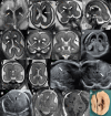
Characteristic features of various malformations of cortical development on ultrasound and T2-weighted 3T MRI with accompanying normal cases. [A(i)] A 26 gestational weeks (GW) fetus with lissencephaly. Transvaginal ultrasound (US), coronal plane, shows bilateral abnormal configuration of the Silvian fissures. Courtesy of Eran Kassif. [B(i)] Transvaginal US, axial plane, 20 GW, focal nodular irregularity of the occipital horn (arrow), indicating periventricular nodular heterotopia in a fetus with agenesis of the corpus callosum (arrow). [C(i)] Transvaginal US, parasagittal plane, 24 GW, abnormal early sulcation, suggesting polymicrogyria (arrow). Note corresponding irregular ventricular border (arrowhead). [A(iiiv)] T2-weighted (T2-w) single-shot FSE, coronal and axial planes of a 32 GW fetus with lissencephaly. A posterior-anterior gradient and absent cerebral convolutions at the level of the parietal and posterior frontal lobes (thick arrows, A) and near flat Sylvian fissures. The gyration is markedly reduced in the frontal and temporal lobes. There are small cysts at the level of the ganglionic eminences [thin arrows, A(iii)]. Note the thick subcortical hypointense band in the posterior frontal and parietal lobes [arrowheads, A(iv)]. [B(ii)] T2-w single-shot fast spin-echo (FSE), axial plane, at 21 GW, shows a minor focal irregularity of the ependymal profile of the left lateral ventricle corresponding to a single periventricular nodular heterotopia (thick arrow). [B(iiiiv)] Corresponding axial and sagittal T2-w single-shot FSE images of the same fetus at 30 GW confirm the same findings (thick arrows). Note that the nodular heterotopia is isointense to the cortical layer. [C(iiiv)] T2-w single-shot FSE, axial, coronal and sagittal planes, at 32 GW, fetus with polymicrogyria due to congenital CMV infection, reveal open Sylvian fissures surrounded by an irregular overfolded surface (arrowheads). Mild-moderate ventriculomegaly and T2 hyperintensity of the periventricular white matter in the parieto-occipital regions. [A(vvii), B(vvii) and C(vvii)] Accompanying normal MR images on the same planes.
Figure 5
Figure 5
Ultrasound and T2-weighted MRI, in vivo, post-mortem and postnatal, in subjects with diagnosed tubulinopathy. (A and B) MRI, axial (A) and coronal (B) planes, at 25 gestational weeks (GW) show asymmetric cerebral hemispheres and ventricles in a fetus with TUBB2A mutation. (C) Transvaginal ultrasound (US) at 29 GW of fetus with a TUBB3 mutation depicts asymmetric dysmorphic anterior horns and asymmetric basal ganglia. Courtesy of Zvi Leibovitz. (D) Autopsy, ventral view of the base of the case in AC at 27 GW and (E) post-mortem T2-weighted (T2-w) MRI, axial plane, display abnormal morphology of the brain base and altered frontotemporal size ratio as well as an abnormal configuration of the olfactory bulbs in TUBB2A mutation. (FH) MRI, coronal and axial planes, of the brain of the fetus’ sister at the age of 5 months show hypointense bands (arrows in F and G) that correlate with disorganized white matter pathways shown on diffusion tensor imaging (arrows in H). (IK) T2-w MRI, axial planes, of a fetus with dysgyria due to a TUBA1A mutation at 24 GW; asymmetric Sylvian fissures with bilateral abnormal sulcation (thick arrows). The horns of the lateral ventricles are asymmetric (black asterisks) with a hook-like appearance. Note the marked asymmetry of the basal ganglia (white asterisks) and accompanying normal cases (LN).
Figure 6
Figure 6
Posterior fossa anomalies. Sagittal (A) and axial (F) T2-weighted (T2-w) images of fetal brain at 23 gestational weeks (GW) demonstrating a retrocerebellar arachnoid cyst (star) with associated mass effect manifested by failed descent of the torcular (A), flattening of the posterior margin of the left cerebellar hemisphere (F) and rightward deviation of the falx cerebelli (arrow, F). Diffuse ventriculomegaly suggests extraventricular obstructive hydrocephalus; however, supratentorial ventriculomegaly may in part relate to aqueductal stenosis cause by a congenitally dysgenetic/thickened lower tectum (arrow, A). Despite 4th ventriculomegaly, the tegmento-vermian angle is within normal limits, a finding that militates against Blake’s pouch cyst. Sagittal (B) and axial (G) T2-w images of the fetal brain at 33 GW show isolated enlargement of the cisterna magna (star, B), without focal mass effect, hydrocephalus or abnormality of the brain parenchyma, consistent with a ‘mega cisterna magna’. Elongation and duplication of the falx cerebelli (arrows, G) is a supportive feature. Sagittal T2-w image (C) of the fetal brain at 22 GW depicts a mildly enlarged tegmento-vermian angle and mild 4th ventriculomegaly (C); the brain is otherwise forming normally. Coronal T2-w image (H) reveals only minimal displacement of the choroid plexus which remains located at the central undersurface of the cerebellum rather than being displaced inferolaterally as is seen in the Dandy-Walker phenotype. Together, these findings are consistent with a Blake’s pouch cyst. Sagittal T2-w image (D) of the fetal brain at 21 GW demonstrates an enlarged tegmento-vermian angle, a significantly obtuse fastigial recess angle and deficiency of the lower vermis. Coronal T2-w image (I) shows inferolateral displacement of the tela choroidea/choroid plexus. Together, these findings are consistent with Dandy-Walker phenotype. Sagittal midline (E) and parasagittal (J) T2-w image of the fetal brain at 23 GW demonstrate deficiency of the lower vermis with normal position of the tela choroidea/choroid plexus, extending superomedially towards the lower vermis (arrow, J), compatible with inferior vermian hypoplasia.
Figure 7
Figure 7
T2-weighted 3T MRI follow-up in a case of diagnosed twin-to-twin transfusion syndrome at the fetal age of 16 gestational weeks. (A) Laser ablation of the twin ‘one’ at 19 gestational weeks (GW) due to polyhydramnios. (B) MRI examination of the twin ‘two’ at 25th GW shows right ventriculomegaly on scan in the sagittal plane. (C) MRI follow up of the twin ‘two’ same week, four days later, shows severe porencephaly (the right hemisphere) in the axial plane. (D) MRI reexamination at 29 GW on scan at coronal plane shows the right hemisphere severe porencephaly and abnormal thickened cortex in the left hemispheric posterior perisylvian, parietal, and occipital regions. (EH) T2-weighted MRI follow-up examination of the same subject at age of seventh postnatal month shows severe right porencephaly and cortical malformations in the left hemisphere.

References

    1. Guerrini R, Dobyns WB. Malformations of cortical development: Clinical features and genetic causes. Lancet Neurol. 2014;13:710–726. - PMC - PubMed
    1. Subramanian L, Calcagnotto ME, Paredes MF. Cortical malformations: Lessons in human brain development. Front Cell Neurosci. 2019;13:576. - PMC - PubMed
    1. Barkovich AJ, Guerrini R, Kuzniecky RI, Jackson GD, Dobyns WB. A developmental and genetic classification for malformations of cortical development: Update 2012. Brain. 2012;135:1348–1369. - PMC - PubMed
    1. Raybaud C, Widjaja E. Development and dysgenesis of the cerebral cortex: Malformations of cortical development. Neuroimaging Clin N Am. 2011;21:483–543. - PubMed
    1. Severino M, Geraldo AF, Utz N, et al. . Definitions and classification of malformations of cortical development: Practical guidelines. Brain. 2020;143:2874–2894. - PMC - PubMed