. 2024 Sep;30(3):171.
doi: 10.3892/mmr.2024.13295. Epub 2024 Jul 26.

HSPB8 attenuates lipopolysaccharide‑mediated acute lung injury in A549 cells by activating mitophagy

Affiliations

HSPB8 attenuates lipopolysaccharide‑mediated acute lung injury in A549 cells by activating mitophagy

Xinjian Zhou et al. Mol Med Rep. 2024 Sep.

Abstract

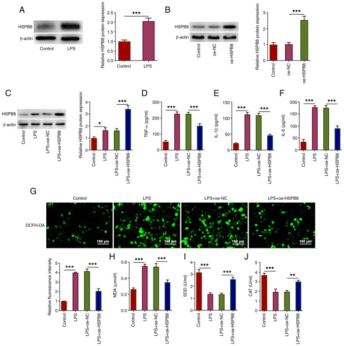
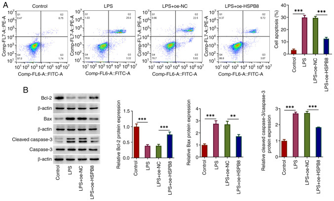
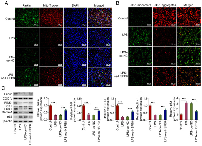
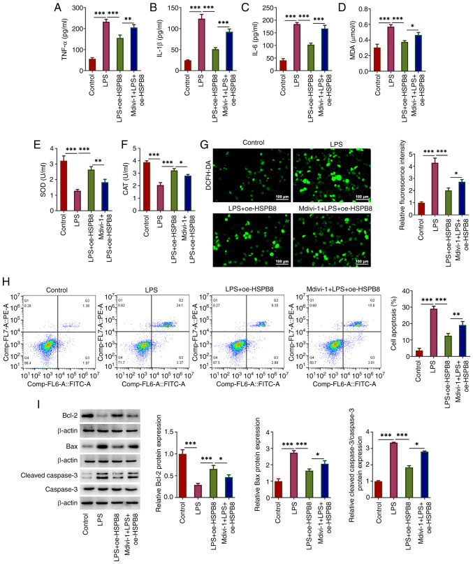
Sepsis is a life‑threatening multiple organ failure disease caused by an uncontrolled inflammatory response and can progress to acute lung injury (ALI). Heat‑shock protein B8 (HSPB8) serves a cytoprotective role in multiple types of diseases; however, to the best of our knowledge, the regulatory role of HSPB8 in sepsis‑induced ALI remains unclear. A549 human alveolar type II epithelial cells were treated with lipopolysaccharide (LPS) for 24 h to simulate a sepsis‑induced ALI model. Cell transfection was performed to overexpress HSPB8, and cells were treated with mitochondrial division inhibitor‑1 (Mdivi‑1) for 2 h before LPS induction to assess the underlying mechanism. Protein expression was evaluated using western blotting and an immunofluorescence assay. Cytokines were examined using ELISA assay kits and antioxidant enzymes were examined using their detection kits. Cell apoptosis was detected using flow cytometry. The mitochondrial membrane potential was detected by JC‑1 staining. HSPB8 was upregulated in A549 cells treated with LPS and HSPB8 overexpression attenuated LPS‑induced inflammatory cytokine levels, oxidative stress and apoptosis in A549 cells. LPS inhibited mitophagy and reduced the mitochondrial membrane potential in A549 cells, which was partly inhibited by HSPB8 overexpression. Furthermore, Mdivi‑1 decreased the inhibitory effect of HSPB8 on the inflammatory response, oxidative stress and apoptosis in LPS‑treated A549 cells. In conclusion, HSPB8 overexpression attenuated the LPS‑mediated inflammatory response, oxidative stress and apoptosis in A549 cells by promoting mitophagy, indicating HSPB8 as a potential therapeutic target in sepsis‑induced ALI.

Keywords: acute lung injury; heat‑shock protein B8; mitophagy; sepsis.


Conflict of interest statement

The authors declare that they have no competing interests.

Figures

Figure 1.
Figure 1.
HSPB8 overexpression attenuates LPS-induced inflammatory cytokine levels and oxidative stress in A549 cells. (A) A549 cells were stimulated by LPS to simulate sepsis-induced acute lung injury. The protein expression levels of HSPB8 were assessed by western blotting. (B) A549 cells were transfected with oe-NC or oe-HSPB8, and the HSPB8 expression was assessed. (C) A549 cells and HSPB8-overexpressing A549 cells were treated with LPS. The protein expression levels of HSPB8 were assessed using western blotting. The concentration of (D) TNF-α, (E) IL-1β and (F) IL-6 in culture medium of A549 cells was detected using ELISAs. (G) Production of intracellular reactive oxygen species was detected by DCFH-DA staining. Scale bar, 100 µm. (H) MDA content, and (I) SOD and (J) CAT activity were examined using commercial kits. *P<0.05, **P<0.01 and ***P<0.001. CAT, catalase; DCFH-DA, 2′-7′-dichlorodihydrofluorescein diacetate; HSP, heat-shock protein; LPS, lipopolysaccharide; MDA malondialdehyde; NC, negative control; oe, overexpression; SOD, superoxide dismutase.
Figure 2.
Figure 2.
HSPB8 overexpression decreases LPS-induced apoptosis in A549 cells. (A) Flow cytometry was used to assess the cell apoptosis rate. (B) Expression levels of apoptosis-related proteins were evaluated using western blotting. **P<0.01 and ***P<0.001. HSP, heat-shock protein; LPS, lipopolysaccharide; NC, negative control; oe, overexpression.
Figure 3.
Figure 3.
HSPB8 overexpression activates mitophagy in LPS-treated A549 cells. (A) Representative double staining with MitoTracker and Parkin. Scale bar, 100 µm. (B) Mitochondrial membrane potential was detected by JC-1 staining. Scale bar, 100 µm. (C) Expression levels of mitophagy-related proteins were examined using western blotting. **P<0.01 and ***P<0.001. HSP, heat-shock protein; LPS, lipopolysaccharide; NC, negative control; oe, overexpression; COX IV, cytochrome c oxidase; PINK1, PTEN-induced kinase 1.
Figure 4.
Figure 4.
Mdivi-1 decreases the inhibitory effects of HSPB8 on the inflammatory response, oxidative stress and apoptosis in LPS-exposed A549 cells. The concentration of (A) TNF-α, (B) IL-1β and (C) IL-6 in the culture medium of A549 cells was detected using ELISAs. (D) MDA content, and (E) SOD and (F) CAT activities were measured using commercial kits. (G) Production of intracellular ROS was detected by DCFH-DA staining. Scale bar, 100 µm. (H) Flow cytometry was performed to investigate the cell apoptosis rate. (I) Expression levels of apoptosis-related proteins were assessed using western blotting. *P<0.05, **P<0.01 and ***P<0.001. CAT, catalase; DCFH-DA, 2′-7′-dichlorodihydrofluorescein diacetate; HSP, heat-shock protein; LPS, lipopolysaccharide; MDA malondialdehyde; Mdivi-1, mitochondrial division inhibitor-1; oe, overexpression; ROS, reactive oxygen species; SOD, superoxide dismutase.

References

    1. Rudd KE, Johnson SC, Agesa KM, Shackelford KA, Tsoi D, Kievlan DR, Colombara DV, Ikuta KS, Kissoon N, Finfer S, et al. Global, regional, and national sepsis incidence and mortality, 1990-2017: Analysis for the global burden of disease study. Lancet. 2020;395:200–211. doi: 10.1016/S0140-6736(19)32989-7. - DOI - PMC - PubMed
    1. Markwart R, Saito H, Harder T, Tomczyk S, Cassini A, Fleischmann-Struzek C, Reichert F, Eckmanns T, Allegranzi B. Epidemiology and burden of sepsis acquired in hospitals and intensive care units: A systematic review and meta-analysis. Intensive Care Med. 2020;46:1536–1551. doi: 10.1007/s00134-020-06106-2. - DOI - PMC - PubMed
    1. Jiang W, Ma C, Bai J, Du X. Macrophage SAMSN1 protects against sepsis-induced acute lung injury in mice. Redox Biol. 2022;56:102432. doi: 10.1016/j.redox.2022.102432. - DOI - PMC - PubMed
    1. Singer M, Deutschman CS, Seymour CW, Shankar-Hari M, Annane D, Bauer M, Bellomo R, Bernard GR, Chiche JD, Coopersmith CM, et al. The third international consensus definitions for sepsis and septic shock (sepsis-3) JAMA. 2016;315:801–810. doi: 10.1001/jama.2016.0287. - DOI - PMC - PubMed
    1. Pham T, Rubenfeld GD. Fifty years of research in ARDS. The epidemiology of acute respiratory distress syndrome. A 50th birthday review. Am J Respir Crit Care Med. 2017;195:860–870. doi: 10.1164/rccm.201609-1773CP. - DOI - PubMed

MeSH terms