. 2024 Jun 21;19(2):183-195.
doi: 10.18502/jovr.v19i2.13278. eCollection 2024 Apr-Jun.

Effect of Pulsed Low-Level Lasers on Adult versus Neonatal Human Retinal Pigment Epithelial Cells: An in-vitro Study

Affiliations

Effect of Pulsed Low-Level Lasers on Adult versus Neonatal Human Retinal Pigment Epithelial Cells: An in-vitro Study

Seyed Mohamadmehdi Moshtaghion et al. J Ophthalmic Vis Res. .

Abstract

Purpose: To investigate the short-term effects of low-level lasers (LLLs; also known as low-power laser therapy) on the structure, genetic, and phenotype of cultured human retinal pigment epithelial (hRPE) cells from both adult and neonatal sources.

Methods: Cultivated adult and neonatal hRPE cells were irradiated with two types of LLL (630 nm and 780 nm), 1 min daily for five consecutive days.

Results: An increase in doubling time was observed in 630 nm-irradiated adult hRPE cells (P = 0.032). The gene expression profile revealed increased expression of retinoid isomerohydrolase RPE65 (RPE65) (P < 0.01 for 630 nm laser, P < 0.001 for 780 nm laser) and nestin (NES) (P < 0.01 for 630 nm laser) in neonatal hRPE cells, upregulation of RPE65 (P < 0.001 for 780 nm laser) and paired box 6 (PAX6) (P < 0.001 for 780 nm laser) genes in adult hRPE cells, and reduced expression of actin alpha 2 (ACTA2) in 780 nm-irradiated adult hRPE cells (P < 0.001). Except the significant increase of α -SMA in 780 nm-irradiated neonatal hRPE cells, no significant change was noted in the expressions of other investigated proteins.

Conclusion: Short-term irradiation of neonatal and adult hRPE cells with LLLs may induce multipotency at the transcriptional level. Irradiation of neonatal hRPE cells with LLLs can be associated with increased risk of myofibroblastic transformation; however, adult hRPE cells irradiated with the 780 nm laser have minimal risk of myofibroblastic differentiation. It seems that the 780 nm laser may be a promising option for future photobiomodulation in retinal degenerations in adults.

Keywords: Human Retinal Pigment Epithelial Cells; Low-level Laser; PAX6; Photobiomodulation; RPE65; Alpha-smooth Muscle Actin.


Conflict of interest statement

None.

Figures
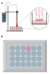
Figure 1
Figure 1
Laser irradiation experimental setup. (a) Illustration of the laser irradiation setup depicting the relative positions of the plate and laser diode. (b) Schematic representation of the plate covered with aluminum foil, featuring a 12 mm aperture designed to minimize scattering effects.
Figure 2
Figure 2
Adult and neonatal hRPE cells irradiated with 630 & 780 nm lasers. Note a mixture of spindle and polygonal morphology at baseline in adult cells irradiated with 630 (a) & 780 nm (b) lasers, and in neonatal cells irradiated with 630 (gr) & 780 nm (hr) lasers. (c) and (i) are the corresponding controls. After 5 days, adult cells irradiated with 630 (d) & 780 nm (e) lasers and their controls (f) still have a mixture morphology. Neonatal cells are predominantly spindle-shaped after a 5-day irradiation with 630 nm laser ( j), and polygonal after a 5-day irradiation with 780 nm laser (k) in comparison to controls (l) that show a mixture morphology. (Scale bar: 100 µm).
Figure 3
Figure 3
Viability, cell proliferation, and gene expression of adult and neonatal hRPE cells irradiated with 630 & 780 nm lasers. (a) MTT shows no significant difference between irradiated and control cells. 630 nm irradiated adult cells show a significant increase of doubling time (P=0.032) compared to control. (b) Note significant upregulation of RPE65 (P < 0.01 for 630 & P < 0.001 for 780 nm) and NES (P < 0.01 for 630 nm) genes in irradiated neonatal cells compared to control. In 780 nm irradiated adult cells, a significant increase of RPE65 and PAX6 (P < 0.001 for both) genes is observed compared to controls. ACTA2 gene in 780 nm irradiated adult cells is significantly downregulated (P < 0.001).
Figure 4
Figure 4
Immune reactivity for fluorescein isothiocyanate (FITC)-conjugated anti-α-smooth muscle actin (α-SMA) antibody in adult hRPE cells irradiated with 630 nm (a) & 780 nm laser (b), and in neonatal hRPE cells irradiated with 630 (d) & 780 nm laser (e) against controls cells (c & f). Propidium iodide-stained cell nuclei are red. Note no significant difference in expression of α-SMA between irradiated and non-irradiate adult cells (g) and significant increase of α-SMA in 780 nm laser-irradiated neonatal cells (P = 0.005) compared to controls, and compared with 630 nm irradiated neonatal cells (P = 0.03) (h).
Figure 5
Figure 5
Immune reactivity for fluorescein isothiocyanate (FITC)-conjugated cytokeratin 8/18 (a), Na+-K+ ATPase (b), and nestin (c) in adult hRPE cells irradiated with 630 (a1,b1,c1) & 780 nm lasers (a2,b2,c2), and in neonatal hRPE cells irradiated with 630 (a5,b5,c5) & 780 nm lasers (a6,b6,c6) against controls (a3,b3,c3 for adult & a7,b7,c7 for neonatal cells). Propidium iodide-stained hRPE cell nuclei are in red. Note no significant alteration in the expression of the proteins between the study groups. (a4,b4,c4 for adult hRPE cells; a8,b8,c8 for neonatal hRPE cells).
Figure 6
Figure 6
Immune reactivity for fluorescein isothiocyanate (FITC)-conjugated ZO-1 (a), RPE65 (b) and PAX-6 (c) in adult hRPE cells irradiated with 630 (a1,b1,c1) & 780 nm lasers (a2,b2,c2), and in neonatal hRPE cells irradiated with 630 (a5,b5,c5) & 780 nm lasers (a6,b6,c6) against controls (a3,b3,c3 for adult & a7,b7,c7 for neonatal cells). Propidium iodide-stained hRPE cell nuclei are in red except for PAX-6 in the nuclei are blue with 4’, 6 -diamidino-2-phenylindole (DAPI) (c series). Note no significant alteration in protein expressions between study groups (a4, b4, c4 for adult & a8, b8, c8 for neonatal cells).
Figure 7
Figure 7
Graphical abstract illustrating the distinct effects of lasers at 630 nm and 780 nm on adult and neonatal hRPE cells.

References

    1. Liu J, Yang L, Wang X, Wang S, Huang Z, Li C, et al. Embryonic stem cell microenvironment enhances proliferation of human retinal pigment epithelium cells by activating the PI3K signaling pathway. Stem Cell Res Ther. 2020;11:411. - PMC - PubMed
    1. Morten la Cour TT. The retinal pigment epithelium. Adv Oral Biol. 2005;10:253–272.
    1. Jin ZB, Gao ML, Deng WL, Wu KC, Sugita S, Mandai M, et al. Stemming retinal regeneration with pluripotent stem cells. Stem Cell Res Ther. 2019;10:38–56. - PubMed
    1. Atmaca-Sonmez P, Li Y, Yamauchi Y, Schanie CL, Ildstad ST, Kaplan HJ, et al. Systemically transferred hematopoietic stem cells home to the subretinal space and express RPE-65 in a mouse model of retinal pigment epithelium damage. Exp Eye Res. 2006;83:1295–1302. - PubMed
    1. Liang CM, Tai MC, Chang YH, Chen YH, Chen CL, Lu DW, et al. Glucosamine inhibits epithelial-to-mesenchymal transition and migration of retinal pigment epithelium cells in culture and morphologic changes in a mouse model of proliferative vitreoretinopathy. Acta Ophthalmol. 2011;89:e505–14. - PubMed

LinkOut - more resources