Review
. 2024 Jun 19;27(7):110296.
doi: 10.1016/j.isci.2024.110296. eCollection 2024 Jul 19.

Medial preoptic circuits governing instinctive social behaviors

Affiliations
Review

Medial preoptic circuits governing instinctive social behaviors

Tamás Láng et al. iScience. .

Abstract

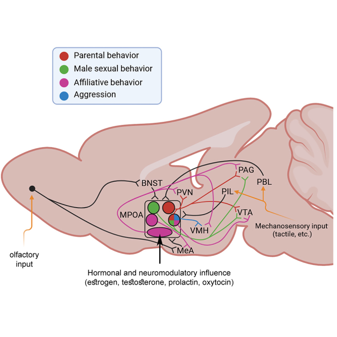
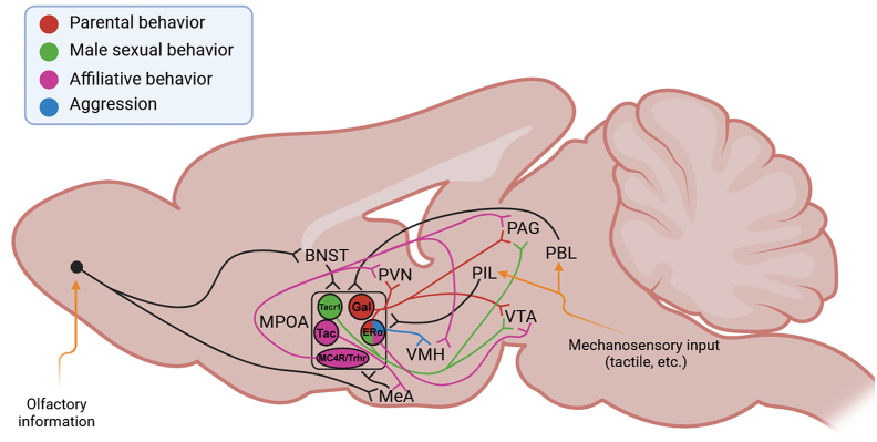
The medial preoptic area (MPOA) has long been implicated in maternal and male sexual behavior. Modern neuroscience methods have begun to reveal the cellular networks responsible, while also implicating the MPOA in other social behaviors, affiliative social touch, and aggression. The social interactions rely on input from conspecifics whose most important modalities in rodents are olfaction and somatosensation. These inputs bypass the cerebral cortex to reach the MPOA to influence the social function. Hormonal inputs also directly act on MPOA neurons. In turn, the MPOA controls social responses via various projections for reward and motor output. The MPOA thus emerges as one of the major brain centers for instinctive social behavior. While key elements of MPOA circuits have been identified, a synthesis of these new data is now provided for further studies to reveal the mechanisms by which the area controls social interactions.

Keywords: Neuroscience; Social sciences.


Conflict of interest statement

The authors declare no competing interests.

Figures

None
Graphical abstract
Figure 1
Figure 1
Schematic diagram shows the preoptic area (POA) and its different subdivisions The POA is located adjacent to the third ventricle. Its periventricular zone contains the periventricular hypothalamic (Pe) and the anteroventral periventricular (AVPV) nuclei. The medial zone includes the medial preoptic nucleus (MPN), the surrounding medial preoptic area (MPOA), the septohypothalamic (SHy), and anterior commissural nuclei (ACN). Functionally, the ventral subdivision of the bed nucleus of stria terminalis (vBNST), which is situated dorsally, is also classified within the medial zone. The MPN and MPA can be further divided into subgroups. The MPN is divided into medial (m), central (c), and lateral (L) regions, while the MPA includes a dorsal area (dMPA). The illustration is based on Paxinos and Franklin’s The Mouse Brain in Stereotaxic Coordinates, and is aligned with a publication on the cellular composition of the preoptic area. The delineation of the medial zone is adapted from a study focusing on this area. Further abbreviations: aca: anterior commissure (anterior part), acp: anterior commissure (posterior), AHA: anterior hypothalamic nucleus (anterior), HDB: nucleus of the horizontal limb of the diagonal band, LA: lateral amygdaloid nucleus, MnPO: median preoptic nucleus, PaAP: paraventricular hypothalamic nucleus (posterior part), PS: parastrial nucleus, SCh: suprachiasmatic nucleus, VLH: ventrolateral hypothalamic nucleus, VLPO: ventrolateral preoptic nucleus, VMPO: ventromedial preoptic nucleus.
Figure 2
Figure 2
A schematic of the parasagittal rodent brain showing the primary projections of glutamatergic and GABAergic preoptic neurons The dashed line indicates the position of bregma. In the preoptic area, vesicular glutamate transporter 2-positive (VGlut2+) glutamatergic neurons are marked by a bold red border on a light green background. Their primary projection sites are denoted by green arrows and light green circles outlined in black. Vesicular GABA transporter-positive (VGAT+) GABAergic neurons in the preoptic region are distinguished by a bold red border and orange coloration. Their projections are indicated by orange arrows and orange circles outlined in black. While vGlut2+ and VGAT+ neurons comprise essentially all preoptic neurons, some cells express both markers. The region enclosed by the dotted line indicates the overlapping population of preoptic vGlut2+ and VGAT+ cells. The major projections of vGlut2+ and VGAT+ neurons include all major projections of the POA. However, distinct cell groups within the POA can have unique projections. Areas with overlapping projections are marked with a gradient. Further abbreviations: AVPV: anteroventral periventricular nucleus, DM: dorsomedial hypothalamic nucleus, LHA: lateral hypothalamic area, PAG: periaqueductal gray, PVN: paraventricular hypothalamic nucleus, SuM: superior mammillary nucleus, VMH: ventromedial hypothalamic nucleus.
Figure 3
Figure 3
Neuronal activation in the preoptic area (POA) of mother mice after exposure to pups compared to that in mother mice separated from their pups Mother mice after 22 h of pup separation with and without receiving their pups back. The coronal sections demonstrate three distinct anatomical levels. The upper panels display representative light microscopic images in a mother mouse demonstrating elevated expression of thec-Fos protein visualized by immunohistochemistry. The lower panels display representative images of a mother mouse separated from her pups, revealing a significantly lower number of activated neurons in the medial preoptic region (MPOA). The region of the medial preoptic nucleus (MPN) and the medial preoptic area (MPA) are bordered by dashed lines. The templates used to indicate the boundaries of the brain regions can be found in the legend of Figure 1. The abbreviations for additional anatomical regions can be found in the legend of Figure 1 and in the list of abbreviations. Scale bar: 200 μm.
Figure 4
Figure 4
The distribution of major socially relevant hormone receptors in the preoptic area (POA) in the mouse brain The figure shows the distribution of prolactin receptor (PRLR), oxytocin receptor (OTR), androgen receptor (AR), and estrogen receptor α (ERα) mRNA obtained by in situ hybridization, as reported by the Allen Mouse Brain Atlas. The selected sections display the expression of each hormone receptor in sexually naive mice, at three different antero-posterior levels located between bregma levels +0.3 and −0.3 mm. PRLR and ERα are particularly abundant in the AVPV and the MPOA. AR has a distribution similar to ERα with lower expression levels in the MPN, while OTR distribution is relatively less pronounced but more widespread in the POA.
Figure 5
Figure 5
Summary of functionally characterized cell types and connections of the medial preoptic area (MPOA) involved in the regulation of social behavior Galanin (Gal)-positive neurons and their projections regulating parental behavior are marked in red. Tachykinin receptor 1 (Tacr1)-expressing neurons and their projections regulating male sexual behavior are shown in green. Several cell populations have been identified that are responsible for the regulation of affiliative behavior, such as melanocortin receptor 4 (MC4R)-, thyrotropin-releasing hormone receptor (Trhr)-, and tachykinin (Tac)-expressing neurons. These cell populations and their projections are shown in purple. While these cell groups likely represent non-overlapping neuronal populations, estrogen receptor α (ERα) is expressed in a large number of MPOA neurons with heterogeneous functions. ERα-expressing neurons have been implicated in all types of social behavior. In cases where a particular projection of ERα neurons has been shown to be involved in a particular type of social behavior, the projections are indicated by the color corresponding to that social behavior. In addition, the blue color indicated the projection from ERα neurons to the ventromedial hypothalamic nucleus (VMH) regulating aggression. Note that the indicated cell groups have projections to several other brain regions that are not indicated due to lack of available functional information. Similarly, additional types of input other than olfactory and mechanosensory are likely but are not indicated in the summary figure due to lack of available knowledge about them. Other abbreviations: BNST: bed nucleus of the stria terminalis, MeA: medial amygdaloid nucleus, PAG: periaqueductal gray, PBL: parabrachial nucleus, PIL: posterior intralaminar thalamic nucleus, PVN: paraventricular hypothalamic nucleus, VTA: ventral tegmental area.
None

Similar articles

Cited by

  • Brain circuits that regulate social behavior.
    Li H, Zhao Z, Jiang S, Wu H. Li H, et al. Mol Psychiatry. 2025 Jul;30(7):3240-3256. doi: 10.1038/s41380-025-03037-6. Epub 2025 Apr 26. Mol Psychiatry. 2025. PMID: 40287553 Free PMC article. Review.

References

    1. Remedios R., Kennedy A., Zelikowsky M., Grewe B.F., Schnitzer M.J., Anderson D.J. Social behaviour shapes hypothalamic neural ensemble representations of conspecific sex. Nature. 2017;550:388–392. doi: 10.1038/nature23885. - DOI - PMC - PubMed
    1. Parise L.F., Joseph Burnett C., Russo S.J. Early life stress and altered social behaviors: A perspective across species. Neurosci. Res. 2023 doi: 10.1016/j.neures.2023.11.005. S0168-0102(23)00200-6. - DOI - PMC - PubMed
    1. Wei D., Talwar V., Lin D. Neural circuits of social behaviors: Innate yet flexible. Neuron. 2021;109:1600–1620. doi: 10.1016/j.neuron.2021.02.012. - DOI - PMC - PubMed
    1. Dickinson S.Y., Kelly D.A., Padilla S.L., Bergan J.F. From Reductionism Toward Integration: Understanding How Social Behavior Emerges From Integrated Circuits. Front. Integr. Neurosci. 2022;16 doi: 10.3389/fnint.2022.862437. - DOI - PMC - PubMed
    1. Mei L., Osakada T., Lin D. Hypothalamic control of innate social behaviors. Science. 2023;382:399–404. doi: 10.1126/science.adh8489. - DOI - PMC - PubMed

LinkOut - more resources