. 2024 Jul 24;10(4):e200173.
doi: 10.1212/NXG.0000000000200173. eCollection 2024 Aug.

Clinicopathologic Characterization of 2 Individuals With TBK1 Variants-1 Novel Splice Variant, 2 Proteinopathies: A Case Series

Affiliations

Clinicopathologic Characterization of 2 Individuals With TBK1 Variants-1 Novel Splice Variant, 2 Proteinopathies: A Case Series

Kimiko Domoto-Reilly et al. Neurol Genet. .

Abstract

Objectives: Here, we report detailed clinicopathologic evaluation of 2 individuals with pathogenic variants in TBK1, including one novel likely pathogenic splice variant. We describe the striking diversity of clinical phenotypes among family members and also the brain and spinal cord neuropathology associated with these 2 distinct TBK1 variants.

Methods: Two individuals with pathogenic variants in TBK1 and their families were clinically characterized, and the probands subsequently underwent extensive postmortem neuropathologic examination of their brains and spinal cords.

Results: Multiple affected individuals within a single family were found to carry a previously unreported c.358+3A>G variant, predicted to alter splicing. Detailed histopathologic evaluation of our 2 TBK1 variant carriers demonstrated distinct TDP-43 pathologic subtypes, but shared argyrophilic grain disease (AGD) tau pathology.

Discussion: Although all pathogenic TBK1 variants are associated with TDP-43 pathology, the clinical and histologic features can be highly variable. Within one family, we describe distinct neurologic presentations which we propose are all caused by a novel c.358+3A>G variant. AGD is typically associated with older age, but it has been described as a copathologic finding in other TBK1 variant carriers and may be a common feature in FTLD-TDP due to TBK1.


Conflict of interest statement

K. Domoto-Reilly reports research funding from NIH U19AG063911; B.J. Distad reports no disclosures relevant to the manuscript; D.E. Miller reports participation on a scientific advisory board at Oxford Nanopore Technologies (ONT), engagement in a research agreement with ONT, and has received travel compensation from ONT; Y.-H. Lin reports no disclosures relevant to the manuscript; D. Ivanick reports no disclosures relevant to the manuscript; A.S. Warren reports no disclosures relevant to the manuscript; S. Jayadev reports no disclosures relevant to the manuscript; C. Latimer reports no disclosures relevant to the manuscript. Go to Neurology.org/NG for full disclosures.

Figures

Figure 1
Figure 1. Family Pedigrees
(A) Family pedigree of case 1, including a sibling and parent with ataxia and a grandparent with presumed amyotrophic lateral sclerosis (ALS). (B) Family pedigree of case 2, including a sibling with autopsy confirmed frontotemporal lobar degeneration. FTD = frontotemporal dementia; MND = motor neuron disease.
Figure 2
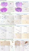
Figure 2. Pathologic TDP-43 and Tau Characteristics
(A) Features of MND. Case 1 showed prominent pallor of the pyramids in the medulla on H&E/LFB stain, but this was not apparent in case 2 (hash-marked outline). Both cases showed pallor of the corticospinal tracts in sections of the spinal cord (asterisks), but this was more severe in case 1. Both cases showed fibrillar pTDP-43 inclusions in upper motor neurons (arrow). The pTDP-43 pathology of lower motor neurons of the anterior horn of the spinal cord included dense cytoplasmic inclusions in case 1 and granular cytoplasmic inclusions in case 2 (arrowhead). (B) Features of FTLD-TDP. Case 1 showed only minimal pTDP-43 pathology in the frontal cortex that was primarily limited to superficial cortical layers and included dense cytoplasm inclusions and rare neurites (circles). The temporal cortex showed more prominent pTDP-43 pathology that included dense cytoplasmic inclusions, some which had donut-like morphology with centrally located nuclei (arrow) and scattered neurites. Although the pathology was more prominent in superficial cortical layers, pTDP-43 pathology was observed in all layers. Case 2 had more severe cortical pTDP-43 pathology in both frontal and temporal cortex compared with case 1. All cortical layers were involved and include both dense and granular cytoplasmic inclusions as well as granular parenchymal pathology. (C) Features of limbic and ventral striatal pathology. Both case 1 and case 2 had significant hippocampal pTDP-43 pathology that included dense paranuclear inclusions in the dentate gyrus. Case 1 had more significant CA1 pathology with dense, tangle-like pTDP-43 inclusions while case 2 had more limited CA1 pathology consisting of scattered granular cytoplasmic pTDP-43 inclusions. The amygdala and ventral striatal pTDP-43 had a similar morphologic appearance to the cortical pTDP-43 in each case. For case 1 this included dense, donut-like cytoplasmic inclusions and for case 2 this was primarily granular cytoplasmic and parenchymal pathology. (D) Features of AGD. Both case 1 and case 2 had the neuropathologic features of argyrophilic grain disease. This includes dendritic swellings (grains) in the neuropil, as seen in the amygdala; perinuclear accentuation of hyperphosphorylated tau (ptau) in CA2 pyramidal neurons of the hippocampus; fuzzy/granular astrocytes in temporal gray matter (circled); and oligodendroglial coiled bodies in white matter (arrowheads).

References

    1. Mackenzie IR, Neumann M, Baborie A, et al. . A harmonized classification system for FTLD-TDP pathology. Acta Neuropathol. 2011;122(1):111-113. doi:10.1007/s00401-011-0845-8 - DOI - PMC - PubMed
    1. Lee EB, Porta S, Baer GM, et al. . Expansion of the classification of FTLD-TDP: distinct pathology associated with rapidly progressive frontotemporal degeneration. Acta Neuropathol. 2017;134(1):65-78. doi:10.1007/s00401-017-1679-9 - DOI - PMC - PubMed
    1. Gijselinck I, Van Mossevelde A, van der Zee J, et al. . Loss of TBK1 is a frequent cause of frontotemporal dementia in a Belgian cohort. Neurology. 2015;85(24):2116-2125. doi:10.1212/WNL.0000000000002220 - DOI - PMC - PubMed
    1. Hirsch-Reinshagen V, Alfaify OA, Hsiung G-YR, et al. . Clinicopathologic correlations in a family with a TBK1 mutation presenting as primary progressive aphasia and primary lateral sclerosis. Amyotroph Lateral Scler Frontotemporal Degener. 2019;20(7-8):568-575. doi:10.1080/21678421.2019.1632347 - DOI - PMC - PubMed
    1. Pinto WBVR, Souza PVS, Farias IB, Badia BML, Chieia MAT, Oliveira ASB. Expanding the spectrum of TBK1-related disorders: adult-onset non-5q spinal muscular atrophy. Revue Neurol (Paris). 2022;187:981-990. doi:10.1590/0004-282X-ANP-2020-0429 - DOI - PubMed

LinkOut - more resources