. 2024 Nov 25;120(14):1768-1779.
doi: 10.1093/cvr/cvae157.

Inhibition of endothelial-to-mesenchymal transition in a large animal preclinical arteriovenous fistula model leads to improved remodelling and reduced stenosis

Affiliations

Inhibition of endothelial-to-mesenchymal transition in a large animal preclinical arteriovenous fistula model leads to improved remodelling and reduced stenosis

Yang Xu et al. Cardiovasc Res. .

Abstract

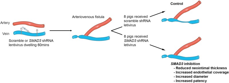
Aims: Vein grafts are used for many indications, including bypass graft surgery and arteriovenous fistula (AVF) formation. However, patency following vein grafting or AVF formation is suboptimal for various reasons, including thrombosis, neointimal hyperplasia, and adverse remodelling. Recently, endothelial-to-mesenchymal transition (EndMT) was found to contribute to neointimal hyperplasia in mouse vein grafts. We aimed to evaluate the clinical potential of inhibiting EndMT and developed the first dedicated preclinical model to study the efficacy of local EndMT inhibition immediately prior to AVF creation.

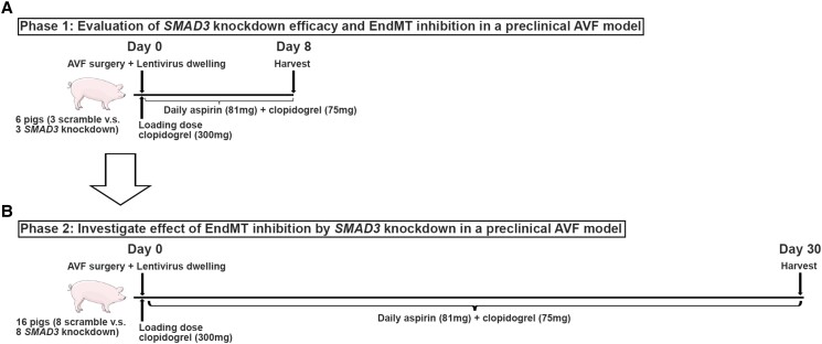
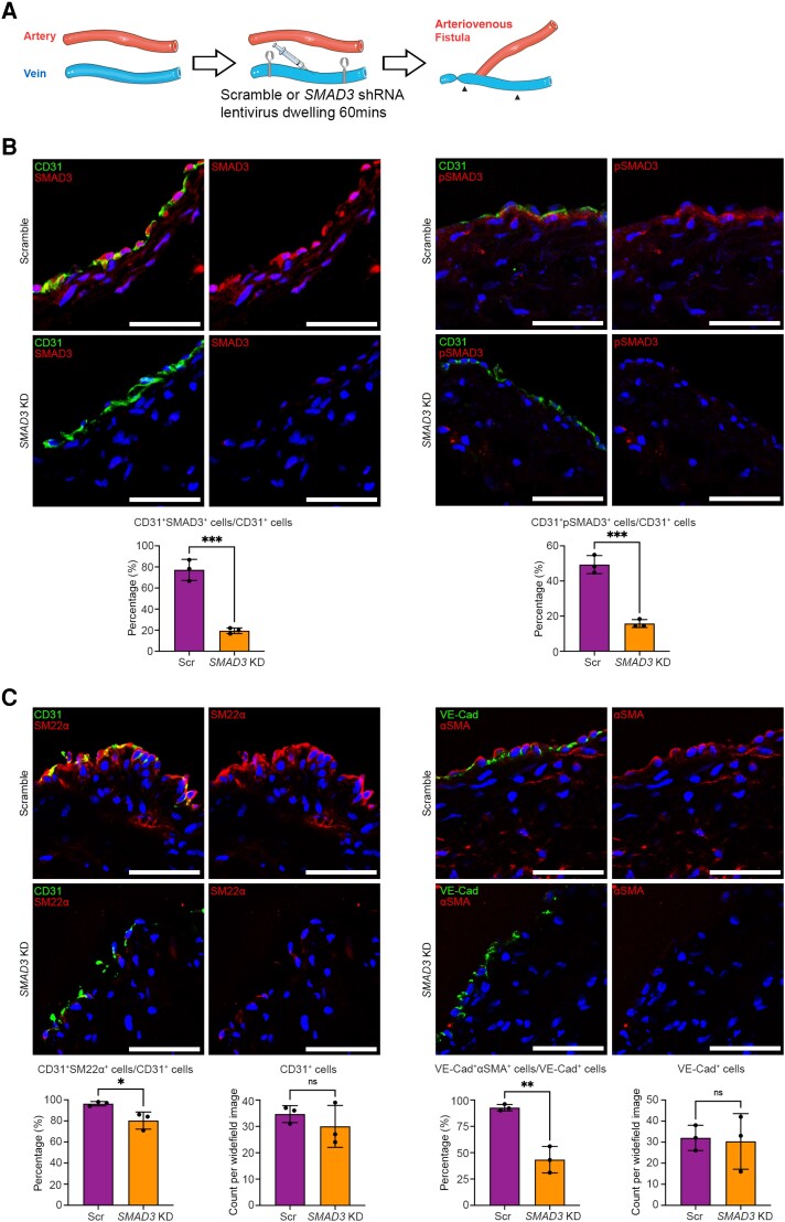
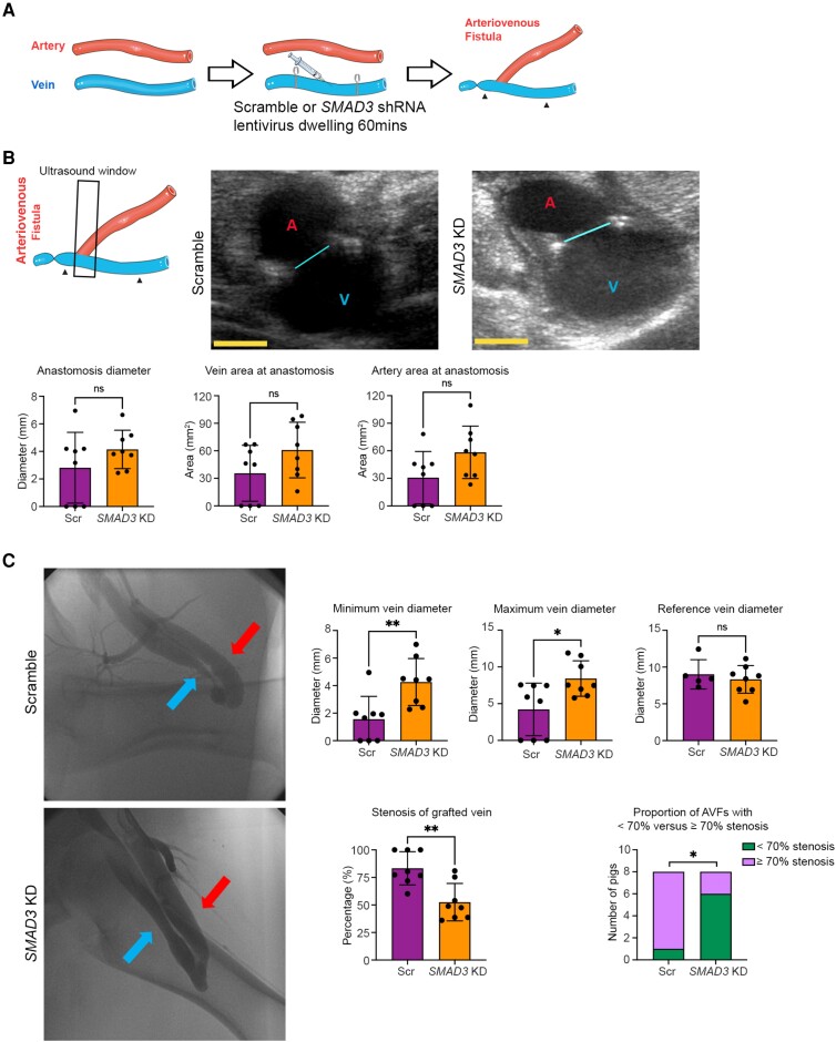
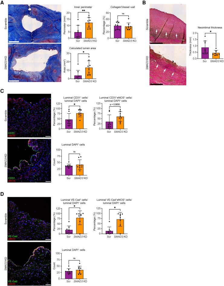
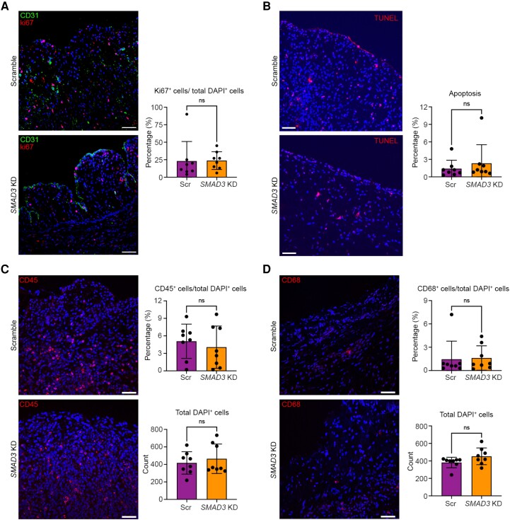
Methods and results: We first undertook pilot studies to optimize the creation of a femoral AVF in pigs and verify that EndMT contributes to neointimal formation. We then developed a method to achieve local in vivo SMAD3 knockdown by dwelling a lentiviral construct containing SMAD3 shRNA in the femoral vein prior to AVF creation. Next, in Phase 1, six pigs were randomized to SMAD3 knockdown or control lentivirus to evaluate the effectiveness of SMAD3 knockdown and EndMT inhibition 8 days after AVF creation. In Phase 2, 16 pigs were randomized to SMAD3 knockdown or control lentivirus and were evaluated to assess longer-term effects on AVF diameter, patency, and related measures at 30 days after AVF creation. In Phase 1, compared with controls, SMAD3 knockdown achieved a 75% reduction in the proportion of CD31+ endothelial cells co-expressing SMAD3 (P < 0.001) and also a significant reduction in the extent of EndMT (P < 0.05). In Phase 2, compared with controls, SMAD3 knockdown was associated with an increase in the minimum diameter of the venous limb of the AVF (1.56 ± 1.66 vs. 4.26 ± 1.71 mm, P < 0.01) and a reduced degree of stenosis (P < 0.01). Consistent with this, neointimal thickness was reduced in the SMAD3 knockdown group (0.88 ± 0.51 vs. 0.45 ± 0.19 mm, P < 0.05). Furthermore, endothelial integrity (the proportion of luminal cells expressing endothelial markers) was improved in the SMAD3 knockdown group (P < 0.05).

Conclusion: EndMT inhibition in a preclinical AVF model by local SMAD3 knockdown using gene therapy led to reduced neointimal hyperplasia, increased endothelialization, and a reduction in the degree of AVF stenosis. This provides important proof of concept to pursue this approach as a clinical strategy to improve the patency of AVFs and other vein grafts.

Keywords: Arteriovenous fistula; Endothelial-to-mesenchymal transition; Neointima; Stenosis; Vein graft.


Conflict of interest statement

Conflict of interest: J.C.K. is named as inventor on provisional patent 63/569 288 filed by Mount Sinai Innovation Partners on 25 March 2024 titled ‘Compositions for reducing SMAD3 expression in a blood vessel and methods of using’. The other authors have no relevant disclosures to declare. The data in this paper were used in a dissertation as partial fulfilment of the requirements for a PhD degree at the Graduate School of Biomedical Sciences at the Icahn School of Medicine at Mount Sinai.

Figures

Graphical Abstract
Graphical Abstract
Inhibition of endothelial-to-mesenchymal transition in a large animal arteriovenous fistula (AVF) model mimicking a human haemodialysis fistula by SMAD3 knockdown with lentiviral gene therapy led to reduced neointimal hyperplasia, increased endothelialization, increased AVF diameter, and potentially to improved AVF patency.
Figure 1
Figure 1
Schematic overview of approach to assessing the efficacy of EndMT inhibition in a preclinical large animal AVF model. (A) Phase 1: Evaluation of SMAD3 knockdown efficacy and EndMT inhibition. The AVF in the right leg was harvested from six pigs, 8 days after AVF creation, where three pigs were randomized to receive lentivirus carrying scramble shRNA and the other three pigs received lentivirus carrying SMAD3 shRNA. (B) Phase 2: Evaluation of the effect and efficacy of EndMT inhibition by SMAD3 knockdown in this pig AVF model. The AVF in the right leg was harvested from 16 pigs, 30 days after AVF creation, where eight pigs were randomized to receive lentivirus carrying scramble shRNA and the other eight pigs received lentivirus carrying SMAD3 shRNA.
Figure 2
Figure 2
Efficacy of SMAD3 knockdown and inhibition of EndMT in preclinical large animal AVF model (Phase 1). (A) Schematic representation of the surgical method including dwelling of lentivirus. Here, in Phase 1, the AVF in the right leg was harvested from six pigs 8 days after AVF creation, where three pigs were randomized to receive lentivirus carrying scramble shRNA (Scr; controls) and three pigs received lentivirus carrying SMAD3 shRNA (SMAD3 KD). Arrowheads indicate the section of vein where the lentivirus was allowed to dwell. (B) Representative immunofluorescence staining and quantitation of SMAD3 and pSMAD3 in endothelial cells 8 days after AVF creation. CD31 is shown in green, SMAD3 or pSMAD3 in red, and DAPI in blue. (C) Representative immunofluorescence staining and quantitation of EndMT and luminal endothelial cell coverage, 8 days after AVF creation. Endothelial markers (CD31 and VE-Cad) are shown in green. Mesenchymal markers (SM22α and αSMA) are in red. DAPI-stained nuclei are in blue. Analyses were performed using unpaired Student’s t-test. All scale bars = 50 µm. *P < 0.05; **P < 0.01; ***P < 0.001. n = 3 pigs per group for all analyses.
Figure 3
Figure 3
Ultrasound and angiographic evaluation of the efficacy of EndMT inhibition by SMAD3 knockdown in preclinical large animal AVF model at 30 days (Phase 2). (A) Schematic representation of the surgical method including dwelling of lentivirus. Here, in Phase 2, the AVF in the right leg was harvested from 16 pigs 30 days after AVF creation, where eight pigs were randomized to receive lentivirus carrying scramble shRNA (Scr; controls) and eight pigs received lentivirus carrying SMAD3 shRNA (SMAD3 KD). Arrowheads indicate the section of vein where the lentivirus was allowed to dwell. (B) Ultrasound measurement at 30 days after AVF creation to assess the surgical anastomotic site and equivalence of AVF creation between groups. The left schematic image represents the orientation of the ultrasound probe during scanning with respect to the AVF. Arrowheads indicate the section of vein where the lentivirus was allowed to dwell. The black frame indicates the ultrasound scanning window. The middle and right panels show representative ultrasound images from scramble (control) and SMAD3 knockdown pigs, respectively. The cyan line indicates the diameter of anastomosis; A in red colour indicates artery; V in blue colour indicates vein. Shown in the panels below are ultrasound quantifications of the anastomosis diameter, vein area and artery area (as acquired at the anastomosis site in the image plane as shown). Yellow scale bar = 5 mm. Anastomosis diameter and artery area were compared using unpaired Student’s t-test, while vein area was compared using a Mann–Whitney test. (C) Angiographic measurement of AVF diameter, stenosis, and patency 30 days after creation. Left panels show representative femoral angiography images. Red arrow indicates artery (arterial limb of AVF) and blue arrow indicates vein (venous limb of AVF). Corresponding diameters and stenosis severity of the venous limb of the AVF are presented on the right. ‘Minimum vein diameter’ represents the minimum diameter of the lentivirus-treated segment of the venous limb of the AVF, ‘maximum vein diameter’ represents the maximal diameter of the lentivirus-treated segment of the venous limb of the AVF, while ‘reference vein diameter’ represents the diameter of the reference vein segment from the adjacent untreated portion of the vein (cranial from the site of lentivirus dwelling). ‘Stenosis of grafted vein’ represents the stenosis of the lentivirus-treated segment of the venous limb of the AVF (determined by comparing the minimum with the reference diameters) presented as either % stenosis or the proportion with stenosis <70% vs. ≥70%. Minimum and maximum vein diameters were compared using Mann–Whitney test. Reference vein diameter and % stenosis of grafted vein were compared using unpaired Student’s t-test. Stenosis of grafted vein (<70% vs. ≥70%) was compared using Fisher’s exact test. *P < 0.05; **P < 0.01; ns, not significant. n = 8 pigs per group for all analyses except ‘reference vein diameter’ in C where n = 5 in the control group only (it was not possible to determine the reference vein diameter in the three occluded AVFs in the control group and therefore these are not presented).
Figure 4
Figure 4
Histologic and immunofluorescence evaluation of the efficacy of EndMT inhibition by SMAD3 knockdown in preclinical large animal AVF model at 30 days (Phase 2). (A) Representative images stained using Masson’s trichrome stain with analyses of inner perimeter, calculated lumen area, and collagen content of the vessel wall. Lumen area was calculated from the inner perimeter (i.e. inner circumference) and assuming the vessel was circular in cross-section. For this panel, all images are from the narrowest portion of the venous limb of the AVF. Scale bar = 1 mm. (B) Representative images stained using EVG stain to identify the inner elastic lamina that demarcates the intima-media boundary (arrows), with quantitation of overall neointimal thickness (from the intima-media boundary to the intima) for each AVF determined by averaging the neointimal thickness measurement from three sites per AVF from a single section. For B, images are from close to the narrowest portion of the venous limb of the AVF (within 1–2 mm). Scale bar = 0.5 mm. (C) Representative immunofluorescence staining for CD31 (green), eNOS (red), and DAPI-stained nuclei (blue) with quantifications. Scale bar = 50 µm. (D) Representative immunofluorescence staining for VE-Cad (green), eNOS (red), and DAPI-stained nuclei (blue) with quantifications. Scale bar = 50 µm. Images in C and D are from the venous limb of the AVF, within 5–10 mm of the narrowest portion. Analyses were performed as follows: (A) inner perimeter with unpaired Student’s t-test and both calculated lumen area and collagen content with Mann–Whitney test; (B) neointimal thickness with unpaired Student’s t-test; (C) CD31+ cells/DAPI+ cells and DAPI+ cells with unpaired Student’s t-test, CD31+eNOS+/DAPI+ cells with Mann–Whitney test; (D) DAPI+ cells with unpaired t-test; other analyses in D with Mann–Whitney test. *P < 0.05; **P < 0.01; ns, not significant. n = 8 pigs per group for all analyses.
Figure 5
Figure 5
Evaluation of the effect of EndMT inhibition by SMAD3 knockdown on cell proliferation, apoptosis, and immune cell infiltration in a preclinical large animal AVF model at 30 days (Phase 2). Images in this figure are from the venous limb of the AVF, within 5–10 mm of the narrowest portion. (A) Assessment of cell proliferation with representative immunofluorescence staining for CD31 (green), Ki67 (red), and DAPI-stained nuclei (blue) with quantifications. (B) Assessment of apoptosis with representative immunofluorescence staining for TUNEL assay (red) and DAPI-stained nuclei (blue) with quantifications. (C) Assessment of immune cell infiltration with representative immunofluorescence staining for CD45 (red) and DAPI-stained nuclei (blue) with quantifications. (D) Assessment of immune cell infiltration with representative immunofluorescence staining for CD68 (red) and DAPI-stained nuclei (blue) with quantifications. All analyses were performed using Mann–Whitney test except for in C; CD45+ cell/total DAPI analysis was performed with unpaired Student’s t-test. All scale bars = 50 µm. ns, not significant. n = 8 pigs per group for all analyses.

Comment in

References

    1. Benjamin EJ, Muntner P, Alonso A, Bittencourt MS, Callaway CW, Carson AP, Chamberlain AM, Chang AR, Cheng S, Das SR, Delling FN, Djousse L, Elkind MSV, Ferguson JF, Fornage M, Jordan LC, Khan SS, Kissela BM, Knutson KL, Kwan TW, Lackland DT, Lewis TT, Lichtman JH, Longenecker CT, Loop MS, Lutsey PL, Martin SS, Matsushita K, Moran AE, Mussolino ME, O'Flaherty M, Pandey A, Perak AM, Rosamond WD, Roth GA, Sampson UKA, Satou GM, Schroeder EB, Shah SH, Spartano NL, Stokes A, Tirschwell DL, Tsao CW, Turakhia MP, VanWagner LB, Wilkins JT, Wong SS, Virani SS, American Heart Association Council on E, Prevention Statistics C and Stroke Statistics S . Heart disease and stroke statistics—2019 update: a report from the American Heart Association. Circulation 2019;139:e56–e528. - PubMed
    1. Foy AJ, Mandrola JM. Heavy heart: the economic burden of heart disease in the United States now and in the future. Prim Care 2018;45:17–24. - PubMed
    1. Lawton JS, Tamis-Holland JE, Bangalore S, Bates ER, Beckie TM, Bischoff JM, Bittl JA, Cohen MG, DiMaio JM, Don CW, Fremes SE, Gaudino MF, Goldberger ZD, Grant MC, Jaswal JB, Kurlansky PA, Mehran R, Metkus TS Jr, Nnacheta LC, Rao SV, Sellke FW, Sharma G, Yong CM, Zwischenberger BA. 2021 ACC/AHA/SCAI guideline for coronary artery revascularization: a report of the American College of Cardiology/American Heart Association joint committee on clinical practice guidelines. Circulation 2022;145:e18–e114. - PubMed
    1. Hess CN, Lopes RD, Gibson CM, Hager R, Wojdyla DM, Englum BR, Mack MJ, Califf RM, Kouchoukos NT, Peterson ED, Alexander JH. Saphenous vein graft failure after coronary artery bypass surgery: insights from PREVENT IV. Circulation 2014;130:1445–1451. - PMC - PubMed
    1. Sabik JF III. Understanding saphenous vein graft patency. Circulation 2011;124:273–275. - PubMed

Publication types

MeSH terms