. 2024 Jul 17;13(14):1208.
doi: 10.3390/cells13141208.

HSV-1 and Cellular miRNAs in CSF-Derived Exosomes as Diagnostically Relevant Biomarkers for Neuroinflammation

Affiliations

HSV-1 and Cellular miRNAs in CSF-Derived Exosomes as Diagnostically Relevant Biomarkers for Neuroinflammation

Christian Scheiber et al. Cells. .

Abstract

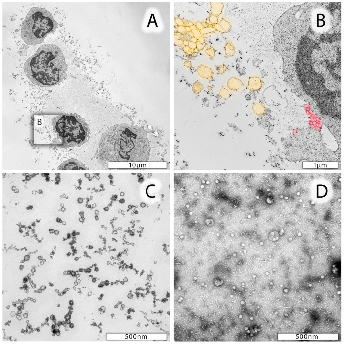
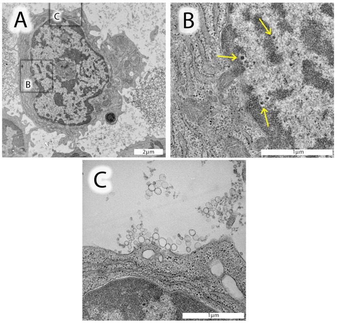
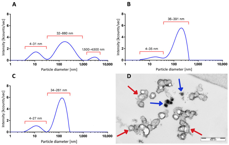
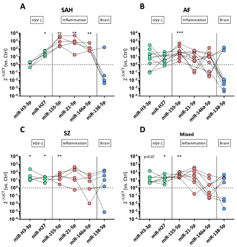
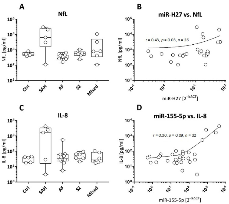
Virus-associated chronic inflammation may contribute to autoimmunity in a number of diseases. In the brain, autoimmune encephalitis appears related to fluctuating reactivation states of neurotropic viruses. In addition, viral miRNAs and proteins can be transmitted via exosomes, which constitute novel but highly relevant mediators of cellular communication. The current study questioned the role of HSV-1-encoded and host-derived miRNAs in cerebrospinal fluid (CSF)-derived exosomes, enriched from stress-induced neuroinflammatory diseases, mainly subarachnoid hemorrhage (SAH), psychiatric disorders (AF and SZ), and various other neuroinflammatory diseases. The results were compared with CSF exosomes from control donors devoid of any neuroinflammatory pathology. Serology proved positive, but variable immunity against herpesviruses in the majority of patients, except controls. Selective ultrastructural examinations identified distinct, herpesvirus-like particles in CSF-derived lymphocytes and monocytes. The likely release of extracellular vesicles and exosomes was most frequently observed from CSF monocytes. The exosomes released were structurally similar to highly purified stem-cell-derived exosomes. Exosomal RNA was quantified for HSV-1-derived miR-H2-3p, miR-H3-3p, miR-H4-3p, miR-H4-5p, miR-H6-3p, miR-H27 and host-derived miR-21-5p, miR-146a-5p, miR-155-5p, and miR-138-5p and correlated with the oxidative stress chemokine IL-8 and the axonal damage marker neurofilament light chain (NfL). Replication-associated miR-H27 correlated with neuronal damage marker NfL, and cell-derived miR-155-5p correlated with oxidative stress marker IL-8. Elevated miR-138-5p targeting HSV-1 latency-associated ICP0 inversely correlated with lower HSV-1 antibodies in CSF. In summary, miR-H27 and miR-155-5p may constitute neuroinflammatory markers for delineating frequent and fluctuating HSV-1 replication and NfL-related axonal damage in addition to the oxidative stress cytokine IL-8 in the brain. Tentatively, HSV-1 remains a relevant pathogen conditioning autoimmune processes and a psychiatric clinical phenotype.

Keywords: CSF exosomes; HSV-1; IL-8; NfL; encephalitis; low-grade inflammation; miRNA; neuronal damage; oxidative stress; psychiatric disease; traumatic brain injury.


Conflict of interest statement

Author Duncan Ross was employed by the company Kimera Labs Inc. The remaining authors declare that the research was conducted in the absence of any commercial or financial relationships that could be construed as a potential conflict of interest.

Figures

Figure 1
Figure 1
Representative electron micrographs of leukocytes from CSF. (A) Lymphocytes and one monocyte in the lower part of the image. Cells are surrounded by a protein matrix and numerous vesicles differing in size. (B) Enlarged area of a lymphocyte marked in (A) with likely extruding microparticles highlighted in yellow and exosomes highlighted in red. (C) Smaller (<40 nm in diameter) particles in CSF specimen which may be due to proteins, oxidized phospholipids, and nucleic acids. (D) Negative staining preparation of mesenchymal stem-cell-derived exosomes.
Figure 2
Figure 2
Electron micrographs of leukocytes from a selected AF patient #15159. (A) Lymphocyte from the CSF with a well-structured, metabolically active nucleus, mitochondria, and endoplasmic reticulum. The nucleus of this lymphocyte contains homo- and heterochromatin and a well-identified nucleolus in the center. Distinct, highly electron-dense spots, structurally similar to herpesvirus nucleocapsids devoid of an envelope, can be identified in the distal area of the heterochromatin. (B) Enlarged insert of the nuclear area (black square in (A)) shows these likely herpesvirus nucleocapsids with a characteristically light halo surrounding the nucleocapsid (yellow arrows). (C) Enlarged insert area from (A) documenting the presence of exosomes (50–120 nm in diameter) close to lymphocyte’s cell surface. In the upper part of this image insert, very small vesicles can be identified, likely corresponding to proteins, oxidized phospholipids, and/or nucleic acid aggregates.
Figure 3
Figure 3
Vesicle size distributions from CSF-derived particles (EVs). (A) DLS-generated size distribution profile of cell-free CSF after step 1 centrifugation from a representative AF patient, with peaks at 12.20 nm, 215.00 nm, and 2790 nm in diameter. (B) Size distribution profile of another patient’s preparation after step 3 ultracentrifugation. Enriched EVs show a major peak at 183.5 nm in diameter and a smaller peak fraction with 16.0 nm in diameter. (C) DLS-generated size distribution pattern of highly purified mesenchymal stem-cell-derived exosomes (XoGloR, Kimeralabs.com) showing a major peak at 124.0 nm and a smaller peak at 11.90 nm in diameter. For vesicle size distribution plots, the relative intensities (kcounts/s, y-axis) were plotted against the log10 values of particles’ diameters (x-axis). (D) Ultrastructure of cryopressure-processed, highly purified mesenchymal stem-cell-derived exosomes (XoGloR, Kimeralabs.com). Preparation contains lipid-bilayer-enclosed vesicles (red arrows) and lipid-bilayer-negative electron-dense aggregates (blue arrows).
Figure 4
Figure 4
miRNA expression profiles of CSF-derived exosomes’ preparations. Log10 fold changes of HSV-1-derived miR-H3-3p and miR-H27 (green symbols), cellular inflammatory miR-155-5p, miR-21-5p, miR-146a-5p (red symbols), and brain-derived miR-138-5p (blue symbols) are shown as dot plots in SAH (A), AF (B), SZ (C), and non-traumatic/non-psychiatric patients (D). Log10 fold changes were calculated using the 2−ΔΔCT method. Control (Ctrl)-derived exosomal preparations served as reference, and the dotted horizontal line (fold change = 1) represents the cut-off for elevated transcripts. For viral miR-H3-3p, n = 5 SAH, n = 10 AF, n = 5 SZ, and n = 7 non-traumatic/non-psychiatric patients were analyzed and normalized against n = 3 Ctrl samples. For viral miR-H27, n = 5 SAH, n = 7 AF, n = 5 SZ, and n = 7 non-traumatic/non-psychiatric patients were analyzed and normalized against n = 4 Ctrl samples. For host miRNAs, n = 5 SAH, n = 11 AF, n = 6 SZ, and n = 8 non-traumatic/non-psychiatric patients were analyzed and normalized against n = 5 Ctrl samples. Wilcoxon rank-sum test was performed to analyze differences against controls (Ctrl). Significant differences are marked by * p ≤ 0.05, ** p ≤ 0.01, and *** p ≤ 0.001.
Figure 5
Figure 5
NfL and IL-8 concentrations in correlation to viral and host miRNAs. (A) CSF NfL concentrations (pg/mL), presented as box plots (median + range), for n = 5 controls (Ctrl) and patients with SAH (n = 5), AF (n = 8), SZ (n = 6), and mixed neuroinflammatory diseases (n = 6). (B) Correlation between n = 26 NfL concentrations (A) and corresponding viral miR-H27 fold changes. (C) CSF IL-8 concentrations (pg/mL), presented as box plots (median + range), for n = 5 controls (Ctrl), and patients with SAH (n = 5), AF (n = 10), SZ (n = 6), and mixed neuroinflammatory diseases (n = 6). (D) Correlation between n = 32 IL-8 concentrations (C) and corresponding host-derived miR-155-5p fold changes. Correlations are presented as scatter plots; r: Spearman correlation coefficient, p: significance, n: number of specimens.

References

    1. Venkatesan A., Benavides D.R. Autoimmune encephalitis and its relation to infection. Curr. Neurol. Neurosci. Rep. 2015;15:3. doi: 10.1007/s11910-015-0529-1. - DOI - PubMed
    1. Amin I., Vajeeha A., Younas S., Afzal S., Shahid M., Nawaz R., Khan M.U., Idrees M. HSV-1 Infection: Role of Viral Proteins and Cellular Receptors. Crit. Rev. Eukaryot. Gene Expr. 2019;29:461–469. doi: 10.1615/CritRevEukaryotGeneExpr.2019025561. - DOI - PubMed
    1. Wilke J.B.H., Hindermann M., Berghoff S.A., Zihsler S., Arinrad S., Ronnenberg A., Barnkothe N., Steixner-Kumar A.A., Röglin S., Stöcker W., et al. Autoantibodies against NMDA receptor 1 modify rather than cause encephalitis. Mol. Psychiatry. 2021;26:7746–7759. doi: 10.1038/s41380-021-01238-3. - DOI - PMC - PubMed
    1. Dawidowski B., Górniak A., Podwalski P., Lebiecka Z., Misiak B., Samochowiec J. The Role of Cytokines in the Pathogenesis of Schizophrenia. J. Clin. Med. 2021;10:3849. doi: 10.3390/jcm10173849. - DOI - PMC - PubMed
    1. Schretlen D.J., Vannorsdall T.D., Winicki J.M., Mushtaq Y., Hikida T., Sawa A., Yolken R.H., Dickerson F.B., Cascella N.G. Neuroanatomic and cognitive abnormalities related to herpes simplex virus type 1 in schizophrenia. Schizophr. Res. 2010;118:224–231. doi: 10.1016/j.schres.2010.01.008. - DOI - PubMed

Publication types

LinkOut - more resources