Review
. 2024 Jul 19;13(14):1221.
doi: 10.3390/cells13141221.

KRAS Mutation Subtypes and Their Association with Other Driver Mutations in Oncogenic Pathways

Affiliations
Review

KRAS Mutation Subtypes and Their Association with Other Driver Mutations in Oncogenic Pathways

Koushik Mondal et al. Cells. .

Abstract

The KRAS mutation stands out as one of the most influential oncogenic mutations, which directly regulates the hallmark features of cancer and interacts with other cancer-causing driver mutations. However, there remains a lack of precise information on their cooccurrence with mutated variants of KRAS and any correlations between KRAS and other driver mutations. To enquire about this issue, we delved into cBioPortal, TCGA, UALCAN, and Uniport studies. We aimed to unravel the complexity of KRAS and its relationships with other driver mutations. We noticed that G12D and G12V are the prevalent mutated variants of KRAS and coexist with the TP53 mutation in PAAD and CRAD, while G12C and G12V coexist with LUAD. We also noticed similar observations in the case of PIK3CA and APC mutations in CRAD. At the transcript level, a positive correlation exists between KRAS and PIK3CA and between APC and KRAS in CRAD. The existence of the co-mutation of KRAS and other driver mutations could influence the signaling pathway in the neoplastic transformation. Moreover, it has immense prognostic and predictive implications, which could help in better therapeutic management to treat cancer.

Keywords: KRAS; cBioPortal; domain; mutation; predictive response; prognostic response; signaling pathway; therapeutic strategy.


Conflict of interest statement

The authors declare no conflict of interest.

Figures

Figure 9
Figure 9
The median overall survival (OS) of KRAS mutant-harboring patients in different types of cancer. The column diagram represents the median OS of KRAS-mutated variants of pancreatic adenocarcinoma, where G12D shows worse survival compared to wild-type (WT) and G12R-mutated variants of KRAS [129] (A). In the case of colorectal adenocarcinoma-mutated KRAS variant at A146 shows poor survival compared to the mutated variants at G12 and Q61 [145] (B). In contrast, in Lung adenocarcinoma, patients harboring G12C mutation show worse survival compared to WT-KRAS (C) [159].
Figure 1
Figure 1
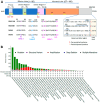
The structure of RAS proteins and prevalence of KRAS mutations in different types of cancer. (A) shows the structure of different isoforms of RAS proteins (KRAS4A, KRAS4B, NRAS, and HRAS). The areas represented with the blue box show the effector lobe (1–86 aa), the orange box as the allosteric lobe (87–166 aa), and the light gray box as a hypervariable region (167–188/189 aa). In the effector lobe, there are P-loop (10–16 aa), Switch Part I (30–38 aa), and Switch Region II (59–76 aa). Yellow boxes (G1–G5) refer to the conserved region responsible for the exchange of guanine nucleotide. The “hotspot” region (12, 13, and Q61) of KRAS remained at the G2 and G3 regions of the protein. The G-domain of RAS family proteins is highly conserved; however, the variability exists at Helix 3, Helix 5, and Loop 8. At Helix 3, Position 95, KRAS is highly conserved and has H (His) for both 4A and 4B, whereas NRAS and HRAS are substituted with L (Leu) and Q (Gln), respectively. In Loop 8, amino acid P (Pro) at Position 120 of KRAS and NRAS is replaced with A (Ala) for HRAS. In comparison, S (Ser) remained conserved at 121 for KRAS4A and 4B. However, S (Ser) is substituted with T (Thr) and A (Ala) in NRAS and HRAS, respectively, at the 121st position of the protein. In Helix 5, Amino Acid Positions 151, 153, 165, and 166 varied among the RAS isoforms. At 151, the R (Arg) of KRAS4A is replaced with G (Gly) of KRAS4B. KRAS4B, NRAS, and HRAS have conserved residues of G (Gly) at Amino Acid Position 151. Next, at 153, amino acid, E (Glu), is conserved among KRAS4A, NRAS, and HRAS, whereas KRAS4B has 153D (153Asp), instead of E (Glu). At Position 165, KRAS4A, NRAS, and HRAS have Q (Gln), whereas Q (Gln) is substituted with K (Lys) in the case of KRAS4B. Both KRAS4A and NRAS have Y (Tyr) at 166th position, while it is occupied with H (His) for KRAS4B and HRAS. There is a conserved motif of CAAX, where cysteine (Cys) is farnesylated, which is denoted as red. Immediately upstream of the CAAX motif in the hypervariable region, there is only about 10–15% homology among four RAS isoforms. In that region, orange is denoted as the site of palmitoylation of Cys residue. KRAS 4B is not palmitoylated; instead, it has a long stretch of lysine as a polybasic domain, while HRAS has two palmitoylated Cys residues. (B) shows the percentages of KRAS mutations in different types of cancer according to the cBioPortal database. TCGA pan-cancer information observed the highest level of KRAS mutation in pancreatic adenocarcinoma, followed by colorectal adenocarcinoma, lung adenocarcinoma, and uterine corpus endometrial carcinoma. Here, green denotes the missense mutations, purple is the structural variant, red is amplification, blue is deep deletion, and grey is multiple alterations.
Figure 2
Figure 2
Activation of KRAS and downstream-signaling pathway. In normal physiological conditions, binding of growth factor to the plasma membrane-associated receptor tyrosine kinase (RTK) induces dimerization and phosphorylation of RTKs. Phosphorylated-RTKs eventually recruit the docking protein, growth factor receptor bound protein-2 (GRB2), and binding with the son of sevenless 1 (SOS1), which itself is a GEF. This activated SOS1, substituting GDP with GTP to KRAS, leading to subsequent conformational changes and activation of downstream factors. The transition from KRAS-GTP to KRAS-GDP, the inactive state, requires GTPase activating protein (GAP). In the activated form of KRAS, it targets downstream effector pathways, namely, (i) RAF/MEK/ERK, (ii) PI3K/Akt/mTOR, and (iii) Ral-GEF/Ral pathway. Mutated KRAS does not need the event of upstream activation. GEF-mediated activation state of KRASmut protein further activates the above-mentioned downstream effector pathways. Blue arrows denote normal physiological conditions, and red arrows indicate the KRAS-mutated state in cancer cells.
Figure 3
Figure 3
Panel of driver mutations in different types of cancer. Following the cBioPortal dataset, figures represented the first 10 driver mutation genes of pancreatic adenocarcinoma (PAAD) (A), colorectal adenocarcinoma (CRAD) (B), and lung adenocarcinoma (LUAD) (C). Data were collected from the cBioPortal of specific types of cancer and associated mutated gene frequencies of that cancer. The bar diagram represents the frequencies of the first 10 highly mutated genes in PAAD (A), CRAD (B), and LUAD (C). The abbreviated form of each gene has been mentioned as follows: Kirsten Ras sarcoma virus (KRAS) is the primary driver mutation gene associated with PAAD, followed by Tumor protein 53 (TP53), SMAD family member 4 (SMAD4), Cylin-dependent kinase inhibitor 2A (CDKN2A), Titin (TTN), Mucin 16 (MUC16), Ring finger protein 43 (RNF 43), Rynodine receptor 1 (RYR1), CUB, and Sushi multiple domain 2 (CSMD2), and Protocadherin-related 15 (PCDH15). In the case of CRAD, Adenomatous polyposis coli (APC) is the primary driver mutation gene, followed by TP53, TTN, KRAS, Phosphatidylinositol-4,5-bisphosphate 3-kinase catalytic subunit alpha (PIK3CA), MUC16, Spectrin repeat containing nuclear envelope protein 1 (SYNE1), FAT atypical cadherin 4 (FAT4), RYR2, and Obscurin (OBSCN). While in LUAD, TP53 is the primary driver mutation gene, followed by TTN, MUC16, CSMD3, RYR2, LDL receptor-related protein 1B (LRP1B), Zinc finger homeobox 4 (ZFHX4), Usherin (USH2A), KRAS, and Xin actin-binding repeat containing 2 (XIRP2).
Figure 4
Figure 4
Mutant variants of KRAS with the coexisting mutations of PIK3CA in colorectal adenocarcinoma (CRAD). In the dot plot (A), mutated KRAS is presented as orange, mutated PIK3CA is presented as pink, the mutations of both KRAS and PIK3CA are presented as red, no mutation is presented as blue, and those not profiled for modification are presented as white. The KRAS-mutated variants are represented as a Doughnut diagram associated with the PIK3CA co-mutation. The high prevalence of mutations of G12D and G12V KRAS is concomitantly associated with PIK3CA mutation in CRAD (A). A positive correlation exists at the mRNA level between the PIK3CA and KRAS (Spearman’s coefficient: 0.37; Pearson’s coefficient: 0.52) (A). The pie diagram represents the first five prevalent PIK3CA mutations coexisting with mutated KRAS variants (B). Among the PIK3CA mutated variants, E545K represents the highest occurrence of co-mutation associated with mutated KRAS, followed by R88Q, C420R, and H1047R (B). Column diagram represents the percentages of KRAS-mutated variants associated with the E545K co-mutation of PIK3CA (C), where G12D remains the prevalent mutation, followed by G13D and others (G12V, A146T, and L19F) (C). Data collected from cBioPortal.
Figure 5
Figure 5
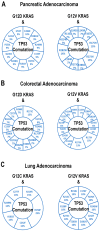
Mutant variants of KRAS with the coexisting mutations of TP53 in pancreatic adenocarcinoma (PAAD) (A), colorectal adenocarcinoma (CRAD (B), and lung adenocarcinoma (C). In the dot plots (AC), mutated KRAS is presented as orange, mutated TP53 is presented as pink, mutation of both KRAS and TP53 are presented as red, no mutation is presented as blue, and not profiled for modification is presented as white. The KRAS-mutated variants are represented as a Doughnut diagram associated with TP53 co-mutation (AC). The high prevalence of mutations of G12D and G12V KRAS is concomitantly associated with TP53 mutations in PAAD (A) and CRAD (B). In contrast, G12V and G12C KRAS mutations are mainly associated with TP53 co-mutation in LUAD (C). Though co-mutation exists, no significant correlation was observed between KRAS and TP53 at the level of mRNA expression (AC). Data collected from cBioPortal.
Figure 6
Figure 6
Co-mutation of TP53 associated with the prevalent KRAS mutations in different types of cancer. Prevalent mutations of KRAS (G12D and G12V) are associated with the missense mutations of TP53 in pancreatic adenocarcinoma (PAAD) (A). R175H, R273H, and R282W are the most frequent mutations of TP53 associated with G12V KRAS in PAAD. Prevalent mutations of KRAS (G12V and G12D) associated with the missense mutations of TP53 in colorectal adenocarcinoma (CRAD) (B). R175H and R273H are the most frequent mutations of TP53 associated with G12V KRAS and R273C is associated with G12D KRAS in CRAD. Prevalent mutations of KRAS (G12V and G12C) are associated with the missense mutations of TP53 in lung adenocarcinoma (LUAD) (C).
Figure 7
Figure 7
Mutant variants of KRAS with the coexisting mutations of APC in colorectal adenocarcinoma (CRAD (A), and Uterine Corpus Endometrial Carcinoma (UCEC) (B). In the dot plots (A,B), mutated KRAS is presented as orange, mutated APC is presented as pink, mutation of both KRAS and APC are presented as red, no mutation is presented as blue, and not profiled for modification is presented as white (A,B). The KRAS-mutated variants associated with APC co-mutation are represented as a Doughnut diagram (A,B). The prevalent KRAS mutations are G12D, G12V, and G13D, which are associated with APC co-mutation in CRAD (A), whereas G12D and G13D are prevalent KRAS mutations associated with APC co-mutation in UCEC (B). Dot plots show the positive correlation of KRAS and APC in CRAD (Spearman’s coefficient: 0.41; Pearson’s coefficient: 0.54) (A), and UCEC (Spearman’s coefficient: 0.60, Pearson’s coefficient: 0.64) (B). Data collected from cBioPortal.
Figure 8
Figure 8
Mutant variants of APC with the coexisting mutations of KRAS in colorectal adenocarcinoma (CRAD) and Uterine corpus endometrial carcinoma (UCEC). Doughnut diagram (A,B) represents the APC-mutated variants associated with KRAS co-mutation. According to cBioPortal, R1450* represents the prevalent APC mutation associated with KRAS co-mutation in CRAD, followed by R213*, E1374*, Q1338*, R232*, and others (A). Along with the APC mutation, the associated co-mutation of KRAS mentioned in (A) in the case of CRAD. The prevalent APC mutations associated with KRAS co-mutation in UCEC are R232*, R1450*, and R1920* (B). Along with the APC mutation, the associated co-mutation of KRAS is mentioned in (B) in the case of UCEC.
Figure 10
Figure 10
Mechanism of inhibition of mutated-KRAS using Sotorasib inhibitor. (A) A mutated form of KRASG12C-GDP inactive state activated by guanine nucleotide exchange factor (GEF) to KRASG12C-GTP active state. KRASG12C-GTP is the cell’s major form due to disrupted GTPase-activating protein (GAP), which converts to KRASG12C-GDP state. The active form of KRASG12C-GTP regulates oncogenic signaling leading to neoplastic transformation. (B) Sotorasib binds explicitly to the KRASG12C-GDP inactive state of the protein and restricts the conversion of the KRASG12C-GTP state leading to the trapping of KRASG12C-GDP. With the restriction of the KRAS-active state, there is inhibition of oncogenic signaling and neoplastic transformation.

References

    1. Poorebrahim M., Abazari M.F., Moradi L., Shahbazi B., Mahmoudi R., Kalhor H., Askari H., Teimoori-Toolabi L. Multi-targeting of K-Ras domains and mutations by peptide and small molecule inhibitors. PLoS Comput. Biol. 2022;18:e1009962. doi: 10.1371/journal.pcbi.1009962. - DOI - PMC - PubMed
    1. Jančík S., Drábek J., Radzioch D., Hajdúch M. Clinical Relevance of KRAS in Human Cancers. J. Biomed. Biotechnol. 2010;2010:150960. doi: 10.1155/2010/150960. - DOI - PMC - PubMed
    1. Tsai F.D., Lopes M.S., Zhou M., Court H., Ponce O., Fiordalisi J.J., Gierut J.J., Cox A.D., Haigis K.M., Philips M.R. K-Ras4A splice variant is widely expressed in cancer and uses a hybrid membrane-targeting motif. Proc. Natl. Acad. Sci. USA. 2015;112:779–784. doi: 10.1073/pnas.1412811112. - DOI - PMC - PubMed
    1. McGrath J.P., Capon D.J., Smith D.H., Chen E.Y., Seeburg P.H., Goeddel D.V., Levinson A.D. Structure and organization of the human Ki-ras proto-oncogene and a related processed pseudogene. Nature. 1983;304:501–506. doi: 10.1038/304501a0. - DOI - PubMed
    1. Karnoub A.E., Weinberg R.A. Ras oncogenes: Split personalities. Nat. Rev. Mol. Cell Biol. 2008;9:517–531. doi: 10.1038/nrm2438. - DOI - PMC - PubMed

MeSH terms

Substances

LinkOut - more resources