. 2024 Jul 20;13(14):1225.
doi: 10.3390/cells13141225.

A Novel Pyrazole Exhibits Potent Anticancer Cytotoxicity via Apoptosis, Cell Cycle Arrest, and the Inhibition of Tubulin Polymerization in Triple-Negative Breast Cancer Cells

Affiliations

A Novel Pyrazole Exhibits Potent Anticancer Cytotoxicity via Apoptosis, Cell Cycle Arrest, and the Inhibition of Tubulin Polymerization in Triple-Negative Breast Cancer Cells

Edgar A Borrego et al. Cells. .

Abstract

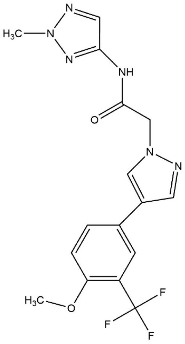
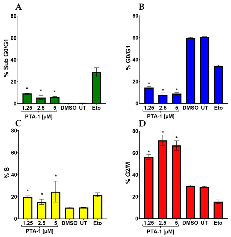
In this study, we screened a chemical library to find potent anticancer compounds that are less cytotoxic to non-cancerous cells. This study revealed that pyrazole PTA-1 is a potent anticancer compound. Additionally, we sought to elucidate its mechanism of action (MOA) in triple-negative breast cancer cells. Cytotoxicity was analyzed with the differential nuclear staining assay (DNS). Additional secondary assays were performed to determine the MOA of the compound. The potential MOA of PTA-1 was assessed using whole RNA sequencing, Connectivity Map (CMap) analysis, in silico docking, confocal microscopy, and biochemical assays. PTA-1 is cytotoxic at a low micromolar range in 17 human cancer cell lines, demonstrating less cytotoxicity to non-cancerous human cells, indicating a favorable selective cytotoxicity index (SCI) for the killing of cancer cells. PTA-1 induced phosphatidylserine externalization, caspase-3/7 activation, and DNA fragmentation in triple-negative breast MDA-MB-231 cells, indicating that it induces apoptosis. Additionally, PTA-1 arrests cells in the S and G2/M phases. Furthermore, gene expression analysis revealed that PTA-1 altered the expression of 730 genes at 24 h (198 upregulated and 532 downregulated). A comparison of these gene signatures with those within CMap indicated a profile similar to that of tubulin inhibitors. Subsequent studies revealed that PTA-1 disrupts microtubule organization and inhibits tubulin polymerization. Our results suggest that PTA-1 is a potent drug with cytotoxicity to various cancer cells, induces apoptosis and cell cycle arrest, and inhibits tubulin polymerization, indicating that PTA-1 is an attractive drug for future clinical cancer treatment.

Keywords: anticancer; apoptosis; cell cycle arrest; drug screening; transcriptome analysis; tubulin inhibition.


Conflict of interest statement

The authors declare no conflicts of interest.

Figures

Figure 1
Figure 1
PTA-1 chemical structure. The chemical structure of 2-{4-[4-methoxy-3-(trifluoromethyl)phenyl]-1H-pyrazol-1-yl}-N-(2-methyl-2H-1,2,3-triazol-4-yl) acetamide, also known as PTA-1.
Figure 2
Figure 2
PTA-1 induces apoptosis. The externalization of PS and the activation of caspase-3/7 indicates that PTA-1 induces apoptosis. (A) Treatment of MDA-MB-231 cells with PTA-1 at concentrations of 10 and 20 μM for 24 h triggers apoptosis, as PS externalization shows. (B) The fluorogenic reagent NucView 488 caspase-3/7 substrate was used to detect the activation of caspase-3/7 via flow cytometry. MDA-MB-231 cells treated with 10 and 20 μM of PTA-1 compound for 7 h significantly induced the activation of caspase-3/7, suggesting apoptosis as the cell death mechanism induced by PTA-1. * indicates p-value lower than 0.01. An average of 3 independent determinations is represented by each bar.
Figure 3
Figure 3
PTA-1 alters the cell cycle by arresting cells in the S and G2/M phases in MDA-MB-231 cells. Exposure of MDA-MB-231 cells to PTA-1 for 72 h causes the alteration of the cell cycle. (A) PTA-1 at 1.25 µM, 2.5 µM, and 5 µM causes significant DNA fragmentation, as measured by the hypodiploid cells that are present in the sub-G0/G1 subpopulations. (B) PTA-1 treatment did not arrest cells in the G0/G1 phase. However, fewer cells in this phase were seen after treatment with the three concentrations of PTA-1. MDA-MB-231 cells exposed to PTA-1 significantly arrested the cells in the S (C) and the G2/M phases (D); nonetheless, the effect was more noticeable in the G2/M phase. Asterisk (*) denotes the p-value being <0.05 as compared to DMSO.
Figure 4
Figure 4
CMap analyses show that PTA-1 treatment results in a gene expression profile similar to that of tubulin inhibitors. MDA-MB-231 cells treated with PTA-1 for 24 h produced a gene expression profile similar to that of known tubulin inhibitors. Results are shown as a heat map in 8 cell lines. The most robust red color means higher positive connections. The top 10 hit compounds based on the tau median score were selected, and all top 10 compounds with gene expression signatures similar to PTA-1 belonged to tubulin inhibitors.
Figure 5
Figure 5
Schrödinger interaction diagrams of PTA-1 and U89 with tubulin. Molecular docking using the Schrödinger software shows the interactions between different molecules and tubulin, and the interactions with the best scores were selected. (A) Diagram shows the PTA-1 hydrogen bond with THR179, ASN247, and ASP249 (red arrows). This interaction resulted in a docking score of −12.345 and an MMGBSA value of −73.78 Kcal/Mol. (B) Compound 89U, used as a positive interaction control, displays a score of −10.926 and an MMGBSA value of −72.89 Kcal/Mol.
Figure 6
Figure 6
PTA-1 disturbs microtubule organization in MDA-MB-231 cells. Cells were stained with an anti-α-tubulin antibody conjugated to Alexa-488 (microtubules), phalloidin–Alexa-568 (microfilaments, polymerized actin), and DAPI (nucleus) and analyzed via confocal microscopy. (A) Four hours of PTA-1 treatment disrupted microtubule organization in MDA-MB231 cells. White arrows indicate empty regions where tubulin is absent. (B) Untreated, (C) DMSO (vehicle), and (D) Paclitaxel (microtubule-stabilizing agent) controls were included.
Figure 7
Figure 7
PTA-1 inhibits tubulin polymerization. Curves of four different treatments for tubulin polymerization. Since absorbance is directly correlated with tubulin polymerization, less absorbance indicates less tubulin polymerization. PTA-1 produced significantly less tubulin polymerization than the vehicle control DMSO at every measurement. PTA-1 and the well-known tubulin polymerization inhibitor vinblastine have similar curves. To compare PTA-1 and DMSO, three independent measurements were taken every 10 min. Asterisk (*) represents a statistical difference of * ≤0.000144 when DMSO and PTA-1 were compared.

References

    1. What Is Cancer?—National Cancer Institute. [(accessed on 31 March 2022)]; Available online: https://www.cancer.gov/about-cancer/understanding/what-is-cancer.
    1. Cancer—Symptoms and Causes—Mayo Clinic. [(accessed on 31 March 2022)]. Available online: https://www.mayoclinic.org/diseases-conditions/cancer/symptoms-causes/sy....
    1. Hanahan D., Weinberg R.A. Hallmarks of Cancer: The next Generation. Cell. 2011;144:646–674. doi: 10.1016/j.cell.2011.02.013. - DOI - PubMed
    1. Wang B., He F., Hu Y., Wang Q., Wang D., Sha Y., Wu J. Cancer Incidence and Mortality and Risk Factors in Member Countries of the “ Belt and Road “ Initiative. BMC Cancer. 2022;22:582. doi: 10.1186/s12885-022-09657-3. - DOI - PMC - PubMed
    1. Siegel R.L., Miller K.D., Wagle N.S., Jemal A. Cancer Statistics, 2023. CA Cancer J. Clin. 2023;73:17–48. doi: 10.3322/caac.21763. - DOI - PubMed

Publication types

MeSH terms

LinkOut - more resources