Review
. 2024 Jul 8;9(7):418.
doi: 10.3390/biomimetics9070418.

Injectable Biomimetic Gels for Biomedical Applications

Affiliations
Review

Injectable Biomimetic Gels for Biomedical Applications

Hossein Omidian et al. Biomimetics (Basel). .

Abstract

Biomimetic gels are synthetic materials designed to mimic the properties and functions of natural biological systems, such as tissues and cellular environments. This manuscript explores the advancements and future directions of injectable biomimetic gels in biomedical applications and highlights the significant potential of hydrogels in wound healing, tissue regeneration, and controlled drug delivery due to their enhanced biocompatibility, multifunctionality, and mechanical properties. Despite these advancements, challenges such as mechanical resilience, controlled degradation rates, and scalable manufacturing remain. This manuscript discusses ongoing research to optimize these properties, develop cost-effective production techniques, and integrate emerging technologies like 3D bioprinting and nanotechnology. Addressing these challenges through collaborative efforts is essential for unlocking the full potential of injectable biomimetic gels in tissue engineering and regenerative medicine.

Keywords: biocompatibility; biomimetic materials; controlled drug delivery; injectable hydrogels; tissue regeneration.


Conflict of interest statement

The authors declare no conflicts of interest.

Figures
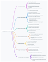
Scheme 1
Scheme 1
Structure–property relationships of injectable biomimetic gels.
Figure 1
Figure 1
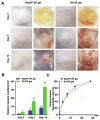
In vivo vascularization and degradation properties of the SV-SF hydrogel after subcutaneous injection into the dorsal side of mice. (A) External images and inside view of volume changes of the SV-SF and NapFF-SF hydrogels, and regenerated blood vessels within implanted gels and surrounding tissues on days 3, 7, and 14 after injection. (B) Summarized data about the relative number of new blood vessels regenerated within implanted gels and surrounding tissues on the dorsal side of mice within 14 days. (C) Summarized data about volume changes of the implanted SV-SF and NapFF-SF hydrogels on the dorsal side of mice within 28 days (*, p < 0.05, **, p < 0.01, ***, p < 0.001); adopted with permission [17].
Figure 2
Figure 2
In vivo wound closure and healing evaluation. (a) Images of the incisions closed by suture, biomedical glue, adhesive hydrogel, and the wound without treatment (set as control). (b) The tensile strength of the healed skin tissues on day 21. (c) Images of H&E staining and Masson’s trichrome staining of the skin tissues after healing for 7 and 21 days. * p  <  0.05. Adopted with permission [2].
Figure 3
Figure 3
In vivo evaluation of bone regeneration. (a) Schematic representation of a rat femoral defect implanted with hydrogel. (b) Representative micro-CT images of the rat femoral bone defects at 5 weeks post-implantation. (c) Morphometric analysis of bone volume/tissue volume and (d) analysis of trabecular bone number. Adopted with permission [14].
Figure 4
Figure 4
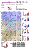
Artificial cartilage formation in vivo. (A) The experimental procedure for injectable SMHs for forming cartilage in vivo. (B) Histochemical staining of chondrocyte-embedded SMH-2 and Matrigel implanted subcutaneously in nude mice. Top, hematoxylin and eosin (H&E) staining; middle, safranin O staining; bottom, collagen II staining. The black line dotted boxes in the upper panel were enlarged in the lower panel. Scale bars, 50 μm. (C) The Young’s moduli of formed artificial cartilages. (D,E) The GAG (D) and collagen II (E) content in chondrocytes cultured within SMH-2 and Matrigel after 6, 8, 10, and 12 weeks. (FH) The relative mRNA levels of Sox9 (F), Col2α1 (G), and Aggrecan (H) in chondrocytes from SMHs after a 12 week implantation in vivo. (I) The protein expression of chondrogenic genes (SOX-9, Col2α1, and Aggrecan) in chondrocytes from SMHs during a 12 week implantation in vivo. * p < 0.05, ** p < 0.01, *** p < 0.001; n = 3 per group per condition. Adopted with permission [69].
Figure 5
Figure 5
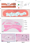
Schematic illustrations and histological images of a regenerative strategy with the use of polyisocyanide (PIC) hydrogel for application in abdominal connective tissue. (A) The application of the hydrogel to the abdominal wound site. (B) PIC-bFGF and PIC-E2, immobilization via azide and the DBCO-linker for the delivery of biomolecules to the tissues. (C) A schematic illustration showing the interaction between the factors and tissue that should trigger regeneration. (D) A histology image of H&E stains confirming the presence of PIC gel between the first (superficial, external oblique) and second (intermediate, internal oblique) muscle layers. (E) A higher magnification of histology images of hydrogel morphology showing a void surrounded by a lining of cells on day 14 and new tissue growth (black arrows) between hydrogel structures (*) on day 42. Adopted with permission [83].
Figure 6
Figure 6
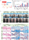
In vivo wound closure and healing performance of the γ-PGA/P-N/CG15 hydrogels in moist and dynamic physiological environment. (A) Schematic representation of the full-thickness rat skin incision models that were created on the relatively dynamic nape, in addition to the static dorsum. (B) Relative tensile strengths of the healed nape and dorsum skins on the 7th and 14th days post-surgery. All results were normalized to the untreated nape and dorsum skins, respectively. (C) Photographs of the full-thickness rat skin incision region on day 0 and the 14th day post-surgery. The red arrows indicate traces of sutures, the red ellipses indicate the unhealed regions of the incisional wounds, and the green lines indicate the healed regions of the incisional wounds. (D) H&E and Masson trichrome staining images of the healed skins on the 14th day post-surgery. Values represent the mean and error bars represent the standard deviation (n = 5). Adopted with permission [94].
Figure 7
Figure 7
A schematic illustration showing (a) the fabrication of GelMA microspheres by microfluidic technology, the synthesis of DMA-MPC polymer by free radical copolymerization, and the design of lubricated GelMA@DMA-MPC microspheres via the dip coating method and (b) a treatment option for osteoarthritis by intra-articular injection of the drug-loaded and lubricated GelMA@DMA-MPC microspheres based on the synergistical intervention of enhanced lubrication and sustained drug release. Adopted with permission [100].

References

    1. Hou M., Wang X., Yue O., Zheng M., Zhang H., Liu X. Development of a multifunctional injectable temperature-sensitive gelatin-based adhesive double-network hydrogel. Biomater. Adv. 2022;134:112556. doi: 10.1016/j.msec.2021.112556. - DOI - PubMed
    1. Liang Y., Xu H., Li Z., Zhangji A., Guo B. Bioinspired Injectable Self-Healing Hydrogel Sealant with Fault-Tolerant and Repeated Thermo-Responsive Adhesion for Sutureless Post-Wound-Closure and Wound Healing. Nano-Micro Lett. 2022;14:185. doi: 10.1007/s40820-022-00928-z. - DOI - PMC - PubMed
    1. Liu Y., Meng H., Konst S., Sarmiento R., Rajachar R., Lee B.P. Injectable dopamine-modified poly(ethylene glycol) nanocomposite hydrogel with enhanced adhesive property and bioactivity. ACS Appl. Mater. Interfaces. 2014;6:16982–16992. doi: 10.1021/am504566v. - DOI - PMC - PubMed
    1. Yan S., Wang W., Li X., Ren J., Yun W., Zhang K., Li G., Yin J. Preparation of mussel-inspired injectable hydrogels based on dual-functionalized alginate with improved adhesive, self-healing, and mechanical properties. J. Mat. Chem. B. 2018;6:6377–6390. doi: 10.1039/C8TB01928B. - DOI - PubMed
    1. Wang R., Liu L., He X., Xia Z., Zhao Z., Xi Z., Yu J., Wang J. Dynamic Crosslinked Injectable Mussel-Inspired Hydrogels with Adhesive, Self-Healing, and Biodegradation Properties. Polymers. 2023;15:13. doi: 10.3390/polym15081876. - DOI - PMC - PubMed

LinkOut - more resources