. 2024 Jun 29;12(7):202.
doi: 10.3390/dj12070202.

A Novel Device for the Evaluation of In Vitro Bacterial Colonization in Membranes for Guided Tissue and Bone Regeneration

Affiliations

A Novel Device for the Evaluation of In Vitro Bacterial Colonization in Membranes for Guided Tissue and Bone Regeneration

Ana Clara Kuerten Gil et al. Dent J (Basel). .

Abstract

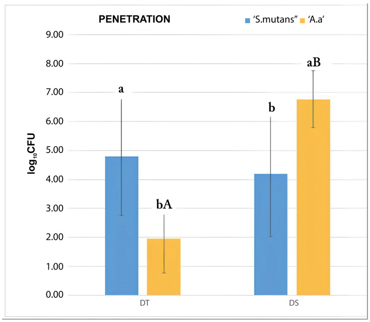
Purpose: To evaluate, in vitro, the efficiency of a novel apparatus to test the adherence and penetration of bacteria on different membranes for guided regeneration. Methodology: To create the 3D device, Computer Aided Design/Computer Aided Manufacturing (CAD/CAM) systems were used. Three types of biomaterials were tested (n = 6): (DT) a collagen membrane; (DS) a polymer membrane; and (LP) a dense polytetrafluoroethylene barrier. The biomaterials were adapted to the apparatuses and challenged with two different monospecies bacterial culture of A. actinomycetemcomitans b and S. mutans. After 2 h, bacterial adherence and penetration were quantified by counting the number of colony-forming units (CFUs). Two specimens from each group were used for image analysis using Confocal Laser Scanning Microscopy. Statistical analysis was performed. Findings: The DS group had a higher adherence of S. mutans compared to A. actinomycetemcomitans b (p = 0.05). There was less adherence of A. actinomycetemcomitans b in the DS group, compared to the LP (p = 0.011) and DT (p < 0.001) groups. Only the membranes allowed penetration, which was blocked by barriers. The DT group allowed a greater penetration of S. mutans to occur compared to A. actinomycetemcomitans b (p = 0.009), which showed a higher penetration into the DS membranes compared to S. mutans (p = 0.016). The penetration of A. actinomycetemcomitans b through DS was higher compared to its penetration through DT and LP (p < 0.01 for both). DT and DS allowed a greater penetration of S. mutans to occur compared to LP, which prevented both bacterial species from penetrating. Conclusion: The apparatus allowed for the settlement and complete sealing of the biomaterials, enabling standardization.

Keywords: 3D; CAD/CAM; bacteria; guided bone regeneration; guided tissue regeneration.


Conflict of interest statement

The authors declare no conflicts of interest.

Figures

Figure 1
Figure 1
(A,B). Methodological apparatus. (A) Virtual design of the methodological apparatus, with unthreaded chambers (elaborated by the authors). (B) Total and individual measurements for each component of the methodological apparatus (elaborated by the authors).
Figure 1
Figure 1
(A,B). Methodological apparatus. (A) Virtual design of the methodological apparatus, with unthreaded chambers (elaborated by the authors). (B) Total and individual measurements for each component of the methodological apparatus (elaborated by the authors).
Figure 2
Figure 2
Bacterial adherence test by CFU counting. Average values from the triplicate analysis. Upper case letters indicate statistical difference between experimental groups and lower-case letters indicate statistical difference between bacterial species, in the groups. DS group showed higher adherence of S. mutans than A. actinomycetemcomitans b and lower adherence of A. actinomycetemcomitans b compared to LP and DT groups. DT group showed higher adherence of S. mutans compared to LP group, whose adherence was the lowest among the groups (elaborated by the authors).
Figure 3
Figure 3
(AC) Viable S. mutans and (DF) A. actinomycetemcomitans cells stained in green and non-viable cells stained in red (Images obtained by Karina Cesca at the Multi-User Laboratory for Studies in Biology and Central Electron Microscopy Laboratory). (A) Balance between viable and non-viable S. mutans cells on DS membrane surface (2500× magnification). (B) Abundant predominance of viable S. mutans cells on DT membrane surface, where no non-viable cells were identified (2500× magnification). (C) Predominance of small proportions of viable S. mutans cells, with no non-viable cells identified on LP barrier (2500× magnification). (D) Predominance of non-viable A. actinomycetemcomitans b cells on DS membrane, with green traces indicating microbial mobility and foci of viable A. actinomycetemcomitans b cells (2500× magnification). (E) Greater amount of viable A. actinomycetemcomitans b cells in DT membrane surface, with small foci of non-viable cells (2500× magnification). (F) Greater amount of non-viable A. actinomycetemcomitans b cells on LP barrier (2500× magnification).
Figure 4
Figure 4
Bacterial penetration test carried out through CFU counting. Average values from the triplicate analysis. Upper-case letters indicate statistical difference between experimental groups and lower-case letters indicate statistical difference between bacterial species in the groups. DT group showed higher penetration of S. mutans compared to A. actinomycetemcomitans b, while A. actinomycetemcomitans b showed higher penetration in DS group compared to S. mutans. Penetration of A. actinomycetemcomitans b across DS group was higher compared to the DT and LP groups. DT and DS groups showed higher penetration of S. mutans compared to LP group, which prevented both bacterial species from penetration (elaborated by the authors).

Similar articles

Cited by

References

    1. Melcher A.H. On the repair potential of periodontal tissues. J. Periodontol. 1976;47:256–260. doi: 10.1902/jop.1976.47.5.256. - DOI - PubMed
    1. Tumedei M., Mourão C.F., D’Agostino S., Dolci M., Di Cosola M., Piattelli A., Lucchese A. Histological and Histomorphometric Effectiveness of Barrier Membranes for Jawbone Regeneration: An Overview of More Than 30 Years’ Experience of Research Results of the Italian Implant Retrieval Center (1988–2020) Appl. Sci. 2021;11:2438. doi: 10.3390/app11052438. - DOI
    1. Soldatos N.K., Stylianou P., Koidou V.P., Angelov M., Yukna R., Romanos G.E. Limitations and options using resorbable versus nonresorbable membranes for successful guided bone regeneration. Quintessence Int. 2017;48:131–147. doi: 10.3290/j.qi.a37133. - DOI - PubMed
    1. Wang H.L., Carroll M.J. Guided bone regeneration using bone grafts and collagen membranes. Quintessence Int. 2001;32:504–515. - PubMed
    1. Gao Y., Wang S., Shi B., Wang Y., Chen Y., Wang X., Lee E.-S., Jiang H.-B. Advances in Modification Methods Based on Biodegradable Membranes in Guided Bone/Tissue Regeneration: A Review. Polymers. 2022;14:871. doi: 10.3390/polym14050871. - DOI - PMC - PubMed

LinkOut - more resources