

Electrophoretic Deposition of Chitosan Coatings on the Porous Titanium Substrate
- PMID: 39057310
- PMCID: PMC11277708
- DOI: 10.3390/jfb15070190
Electrophoretic Deposition of Chitosan Coatings on the Porous Titanium Substrate
Abstract
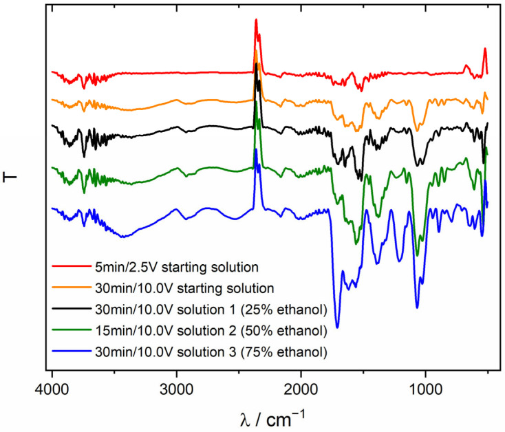
Medicine is looking for solutions to help implant patients recover more smoothly. The porous implants promote osteointegration, thereby providing better stabilization. Introducing porosity into metallic implants enhances their biocompatibility and facilitates osteointegration. The introduction of porosity is also associated with a reduction in Young's modulus, which reduces the risk of tissue outgrowth around the implant. However, the risk of chronic inflammation remains a concern, necessitating the development of coatings to mitigate adverse reactions. An interesting biomaterial for such modifications is chitosan, which has antimicrobial, antifungal, and osteointegration properties. In the present work, a porous titanium biomaterial was obtained by powder metallurgy, and electrophoretic deposition of chitosan coatings was used to modify its surface. This study investigated the influence of ethanol content in the deposition solution on the quality of chitosan coatings. The EPD process facilitates the control of coating thickness and morphology, with higher voltages resulting in thicker coatings and increased pore formation. Ethanol concentration in the solution affects coating quality, with higher concentrations leading to cracking and peeling. Optimal coating conditions (30 min/10 V) yield high-quality coatings, demonstrating excellent cell viability and negligible cytotoxicity. The GIXD and ATR-FTIR analysis confirmed the presence of deposited chitosan coatings on Ti substrates. The microstructure of the chitosan coatings was examined by scanning electron microscopy. Biological tests showed no cytotoxicity of the obtained materials, which allows for further research and the possibility of their use in medicine. In conclusion, EPD offers a viable method for producing chitosan-based coatings with controlled properties for biomedical applications, ensuring enhanced patient outcomes and implant performance.
Keywords: chitosan; electrophoretic deposition; porous titanium.
Conflict of interest statement
The authors declare no conflicts of interest.
Figures


Similar articles
-
Chitosan-Nanogold and Chitosan-Nanozinc Electrodeposited Coatings for Biomedical Applications.J Biomed Mater Res B Appl Biomater. 2025 Apr;113(4):e35571. doi: 10.1002/jbm.b.35571. J Biomed Mater Res B Appl Biomater. 2025. PMID: 40167587
-
Electrophoretic deposition of chitosan coatings on the Ti15Mo biomedical alloy from a citric acid solution.RSC Adv. 2020 Apr 1;10(23):13386-13393. doi: 10.1039/d0ra01481h. eCollection 2020 Apr 1. RSC Adv. 2020. PMID: 35492977 Free PMC article.
-
Highly Efficient Electrophoretic Deposition of Durable, Corrosion-Resistant Chitosan-PEG Composites on Metallic Implants.ACS Appl Mater Interfaces. 2025 Mar 5;17(9):14460-14476. doi: 10.1021/acsami.4c18443. Epub 2025 Feb 18. ACS Appl Mater Interfaces. 2025. PMID: 39964796
-
Surface functionalization of chitosan as a coating material for orthopaedic applications: A comprehensive review.Carbohydr Polym. 2021 Mar 1;255:117487. doi: 10.1016/j.carbpol.2020.117487. Epub 2020 Dec 5. Carbohydr Polym. 2021. PMID: 33436247
-
Metallization of Porous Polyethylene Using a Wire-Arc Spray Process for Heat Transfer Applications.J Therm Spray Technol. 2021;30(1-2):145-156. doi: 10.1007/s11666-020-01119-1. Epub 2021 Jan 3. J Therm Spray Technol. 2021. PMID: 38624489 Free PMC article. Review.
References
LinkOut - more resources
Full Text Sources
Miscellaneous
