

Streamlining Skin Regeneration: A Ready-To-Use Silk Bilayer Wound Dressing
- PMID: 39057462
- PMCID: PMC11276312
- DOI: 10.3390/gels10070439
Streamlining Skin Regeneration: A Ready-To-Use Silk Bilayer Wound Dressing
Abstract
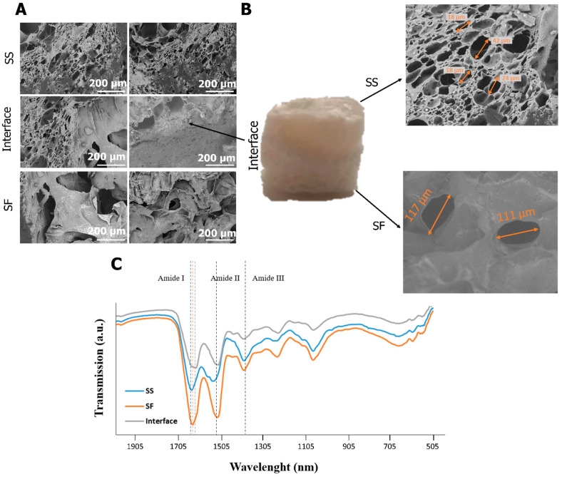
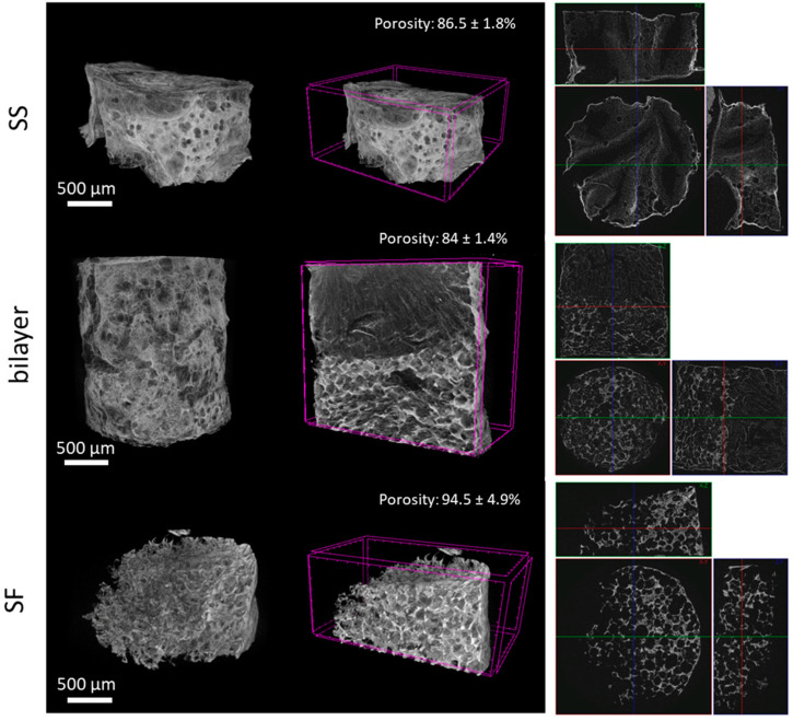
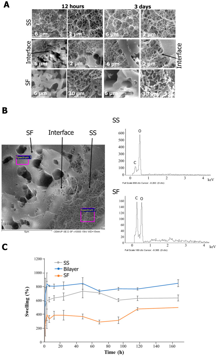
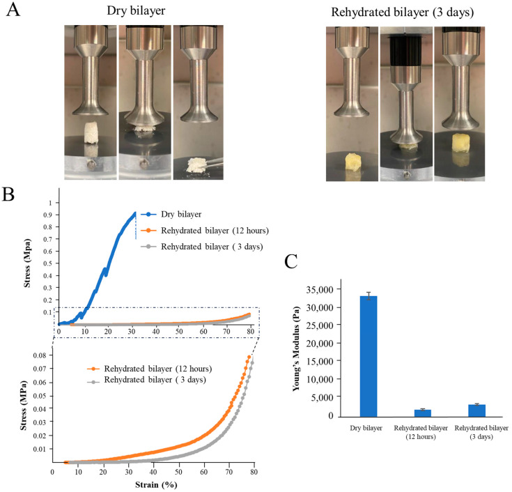
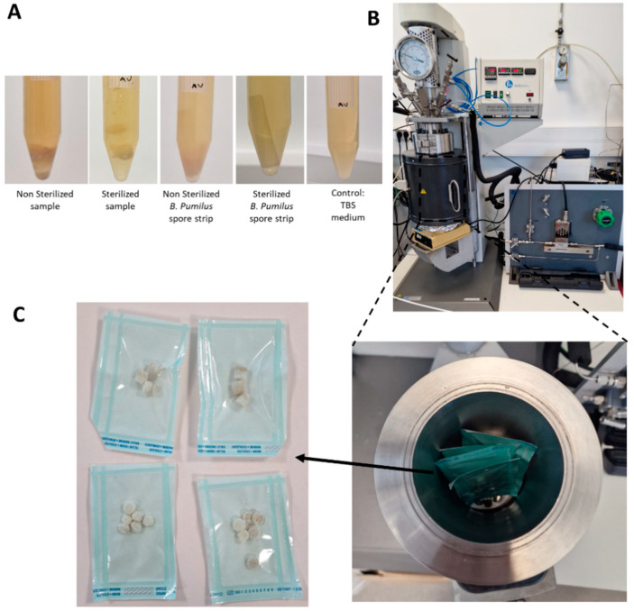
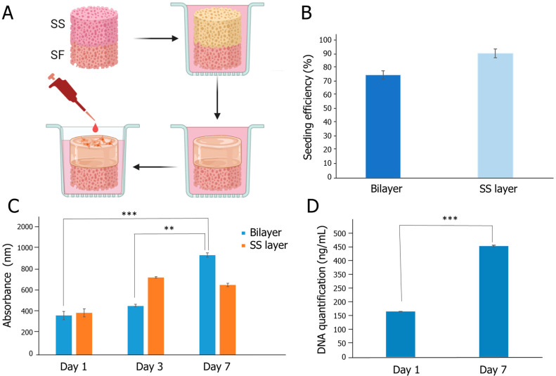
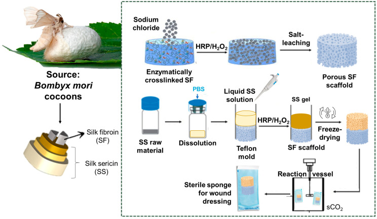
Silk proteins have been highlighted in the past decade for tissue engineering (TE) and skin regeneration due to their biocompatibility, biodegradability, and exceptional mechanical properties. While silk fibroin (SF) has high structural and mechanical stability with high potential as an external protective layer, traditionally discarded sericin (SS) has shown great potential as a natural-based hydrogel, promoting cell-cell interactions, making it an ideal material for direct wound contact. In this context, the present study proposes a new wound dressing approach by developing an SS/SF bilayer construct for full-thickness exudative wounds. The processing methodology implemented included an innovation element and the cryopreservation of the SS intrinsic secondary structure, followed by rehydration to produce a hydrogel layer, which was integrated with a salt-leached SF scaffold to produce a bilayer structure. In addition, a sterilization protocol was developed using supercritical technology (sCO2) to allow an industrial scale-up. The resulting bilayer material presented high porosity (>85%) and interconnectivity while promoting cell adhesion, proliferation, and infiltration of human dermal fibroblasts (HDFs). SS and SF exhibit distinct secondary structures, pore sizes, and swelling properties, opening new possibilities for dual-phased systems that accommodate the different needs of a wound during the healing process. The innovative SS hydrogel layer highlights the transformative potential of the proposed bilayer system for biomedical therapeutics and TE, offering insights into novel wound dressing fabrication.
Keywords: bilayer; silk fibroin; silk sericin; wound dressing.
Conflict of interest statement
The authors declare no conflicts of interest.
Figures

Similar articles
-
Bilayer silk fibroin/sodium alginate scaffold promotes vascularization and advances inflammation stage in full-thickness wound.Biofabrication. 2022 Jun 10;14(3). doi: 10.1088/1758-5090/ac73b7. Biofabrication. 2022. PMID: 35617935
-
Design and performance of sericin/poly(vinyl alcohol) hydrogel as a drug delivery carrier for potential wound dressing application.Mater Sci Eng C Mater Biol Appl. 2019 Aug;101:341-351. doi: 10.1016/j.msec.2019.03.111. Epub 2019 Mar 29. Mater Sci Eng C Mater Biol Appl. 2019. PMID: 31029327
-
Combinatory approach for developing silk fibroin scaffolds for cartilage regeneration.Acta Biomater. 2018 May;72:167-181. doi: 10.1016/j.actbio.2018.03.047. Epub 2018 Apr 5. Acta Biomater. 2018. PMID: 29626700
-
Silk biomaterials in wound healing and skin regeneration therapeutics: From bench to bedside.Acta Biomater. 2020 Feb;103:24-51. doi: 10.1016/j.actbio.2019.11.050. Epub 2019 Dec 2. Acta Biomater. 2020. PMID: 31805409 Review.
-
Silk-Based Biomaterials in Cutaneous Wound Healing: A Systematic Review.Adv Skin Wound Care. 2018 Dec;31(12):565-573. doi: 10.1097/01.ASW.0000546233.35130.a9. Adv Skin Wound Care. 2018. PMID: 30475285
Cited by
-
Innovative Processing and Sterilization Techniques to Unlock the Potential of Silk Sericin for Biomedical Applications.Gels. 2025 Feb 6;11(2):114. doi: 10.3390/gels11020114. Gels. 2025. PMID: 39996657 Free PMC article.
-
Flavonoids regulating NLRP3 inflammasome: a promising approach in alleviating diabetic peripheral neuropathy.Inflammopharmacology. 2025 May;33(5):2231-2262. doi: 10.1007/s10787-025-01729-7. Epub 2025 Apr 9. Inflammopharmacology. 2025. PMID: 40205269 Free PMC article. Review.
References
-
- Martins-Green M. Cutaneous chronic wounds: A worldwide silent epidemic. Open Access Gov. 2023;38:51–53. doi: 10.56367/OAG-038-10647. - DOI
-
- Schlottmann F., Bucan V., Vogt P.M., Krezdorn N. A short history of skin grafting in burns: From the gold standard of autologous skin grafting to the possibilities of allogeneic skin grafting with immunomodulatory approaches. Medicina. 2021;57:225. doi: 10.3390/medicina57030225. - DOI - PMC - PubMed
-
- Gefen A., Alves P., Beeckman D., Lázaro-Martínez J.L., Lev-Tov H., Najafi B., Swanson T., Woo K. Mechanical and contact characteristics of foam materials within wound dressings: Theoretical and practical considerations in treatment. Int. Wound J. 2023;20:1960–1978. doi: 10.1111/iwj.14056. - DOI - PMC - PubMed
-
- Kamińska M.S., Cybulska A.M., Skonieczna-Żydecka K., Augustyniuk K., Grochans E., Karakiewicz B. Effectiveness of Hydrocolloid Dressings for Treating Pressure Ulcers in Adult Patients: A Systematic Review and Meta-Analysis. Int. J. Environ. Res. Public Health. 2020;17:7881. doi: 10.3390/ijerph17217881. - DOI - PMC - PubMed
Grants and funding
- UIDB/50016/2020/Foundation for Science and Technology
- LA/P/0045/2020 (ALiCE), UIDB/00511/2020 and UIDP/00511/2020 (LEPABE)/FCT/MCTES (PIDDAC)
- (0072_IBEROS_MAIS_1_E, Interreg-POCTEP 2021-2027)/IBEROS+
- 2020.08683.BD]/through FCT (Foundation for Science and Technology)
- BE@T-Bioeconomy for Textiles and Apparel, investment TC-C12-i01/Sustainable Bioeconomy, PRR
LinkOut - more resources
Full Text Sources
Molecular Biology Databases
Miscellaneous
