

Congenital Anomalies in American Crocodile (Crocodylus acutus, Cuvier, 1807) Embryos from a Farm Breeder in Colombia
- PMID: 39058001
- PMCID: PMC11281568
- DOI: 10.3390/vetsci11070317
Congenital Anomalies in American Crocodile (Crocodylus acutus, Cuvier, 1807) Embryos from a Farm Breeder in Colombia
Abstract
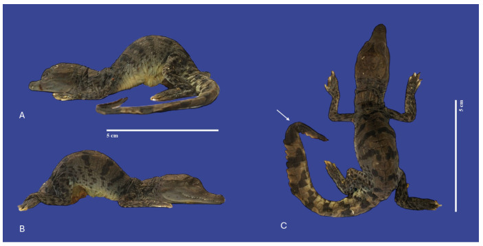
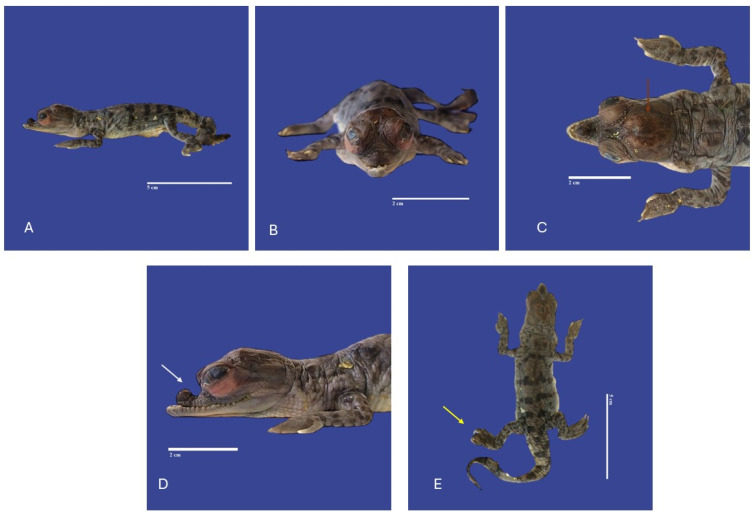
The American crocodile (Crocodylus acutus, Cuvier, 1807) (Class Reptilia, Family Crocodylidae) is a crocodile species inhabiting the Neotropics. Congenital defects have been described in almost every vertebrate group. In crocodiles, teratology alterations have been described in captive animals (pets, zoos, farms) such as Crocodylus niloticus or Gavialis gangeticus. The present study aimed to characterize congenital malformations of C. acutus from a farm in Lomas de Matunilla, Ballestas, Bolívar, Colombia. A total of 550 unhatched eggs were examined after embryo death. A total of 61 embryos presented malformations, with 42 different types of anomalies observed. Limb and tail malformations (29%) were the most common malformations observed. Several malformations, such as cephalothoracopagus, thoracopagus, sternopagus, xiphopagus twins, campylorrachis scoliosa, and acrania, were documented in crocodiles for the first time. Research in teratology enhances our understanding of crocodile biology. It plays a role in their conservation and management, thus helping to ensure the long-term viability of these species in their natural habitats.
Keywords: Crocodylus acutus; caimán aguja; embryos; malformations; teratology.
Conflict of interest statement
The authors declare no conflicts of interest.
Figures


Similar articles
-
Mitochondrial DNA analysis reveals hidden genetic diversity in captive populations of the threatened American crocodile (Crocodylus acutus) in Colombia.Ecol Evol. 2015 Jan;5(1):130-40. doi: 10.1002/ece3.1307. Epub 2014 Dec 8. Ecol Evol. 2015. PMID: 25628870 Free PMC article.
-
Genetic evidence supports a distinct lineage of American crocodile (Crocodylus acutus) in the Greater Antilles.PeerJ. 2018 Nov 12;6:e5836. doi: 10.7717/peerj.5836. eCollection 2018. PeerJ. 2018. PMID: 30473930 Free PMC article.
-
Population assessment of the American crocodile, Crocodylus acutus (Crocodilia: Crocodylidae) on the Pacific coast of Costa Rica.Rev Biol Trop. 2012 Dec;60(4):1889-901. doi: 10.15517/rbt.v60i4.2188. Rev Biol Trop. 2012. PMID: 23342536
-
Similarity of Crocodilian and Avian Lungs Indicates Unidirectional Flow Is Ancestral for Archosaurs.Integr Comp Biol. 2015 Dec;55(6):962-71. doi: 10.1093/icb/icv078. Epub 2015 Jul 3. Integr Comp Biol. 2015. PMID: 26141868 Review.
-
Ice Age effects on genetic divergence of the American crocodile (Crocodylus acutus) in Panama: reconstructing limits of gene flow and environmental ranges: a reply to O'Dea et al.Evolution. 2023 Jan 23;77(1):329-334. doi: 10.1093/evolut/qpac006. Evolution. 2023. PMID: 36689236 Review.
References
-
- American Crocodile (Crocodylus acutus) [(accessed on 30 December 2023)]. Available online: https://www.inaturalist.org/taxa/26085-Crocodylus-acutus.
-
- Rainwater K.L., Wiederhold N.P., Sutton D.A., Garner M.M., Maguire C., Sanders C., Gibas C., Cano J.F., Guarro J., Stchigel A.M. Novel Paranannizziopsis Species in a Wagler’s Viper (Tropidolaemus wagleri), Tentacled Snakes (Erpeton tentaculatum), and a Rhinoceros Snake (Rhynchophis boulengeri) in a Zoological Collection. Med. Mycol. 2019;57:825–832. doi: 10.1093/mmy/myy134. - DOI - PubMed
-
- Ellis T.M. Tolerance of Sea Water by the American Crocodile, Crocodylus acutus. J. Herpetol. 1981;15:187–192. doi: 10.2307/1563379. - DOI
-
- Ogden J.C. Status and Nesting Biology of the American Crocodile, Crocodylus acutus, (Reptilia, Crocodilidae) in Florida. J. Herpetol. 1978;12:183–196. doi: 10.2307/1563406. - DOI
Grants and funding
LinkOut - more resources
Full Text Sources
