. 2024 Jun 28;9(7):145.
doi: 10.3390/tropicalmed9070145.

Experimental Granulomatous Amebic Encephalitis Caused by Acanthamoeba castellanii

Affiliations

Experimental Granulomatous Amebic Encephalitis Caused by Acanthamoeba castellanii

Samuel da Luz Borges et al. Trop Med Infect Dis. .

Abstract

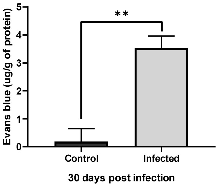
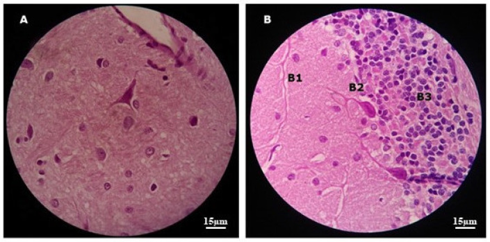
Acanthamoeba genus can affect humans with diseases such as granulomatous amebic encephalitis (GAE), a highly lethal neuroinfection. Several aspects of the disease still need to be elucidated. Animal models of GAE have advanced our knowledge of the disease. This work tested Wistar rats (Rattus norvegicus albinus) as an animal model of GAE. For this, 32 animals were infected with 1 × 10⁶A. castellanii trophozoites of the T4 genotype. Ameba recovery tests were carried out using agar plates, vascular extravasation assays, behavioral tests, and histopathological technique with H/E staining. Data were subjected to linear regression analysis, one-way ANOVA, and Tukey's test, performed in the GraphPad Prism® 8.0 program, with a significance level of p < 0.05. The results revealed the efficiency of the model. Amebae were recovered from the liver, lungs, and brain of infected animals, and there were significant encephalic vascular extravasations and behavioral changes in these animals, but not in the control animals. However, not all infected animals showed positive histopathology for the analyzed organs. Nervous tissues were the least affected, demonstrating the role of the BBB in the defense of the CNS. Supported by the demonstrated evidence, we confirm the difficulties and the feasibilities of using rats as an animal model of GAE.

Keywords: Acanthamoeba; brain; diagnostic; encephalitis; experimental infection; rat.


Conflict of interest statement

The authors declare no conflicts of interest.

Figures

Figure 1
Figure 1
Anatomopathological analysis of the liver and lungs of experimental animals. Images (A,B) show several liver and lung lesions (arrows) compatible with T4r infection. Images (C,D) show these organs preserved in control animals.
Figure 2
Figure 2
Anatomopathological and histopathological images of the liver of an infected rat. Image (A) shows an animal undergoing surgery with liver lesions (arrow) compatible with A. castellanii infection. Image (B) shows a histological section with an overview of one of the liver lesions showing three well-defined areas (circle), at a final magnification of 32×. Image (C) shows these three areas divided into (C1) which represents the peripheral area and shows acidophilic hepatocytes with hyperchromatic nuclei, in addition to mild steatosis. The arrow points to an inflammatory focus. (C2) is the intermediate area characterized by a halo of fibrosis, and (C3) represents the central area of the lesion, characterized by the presence of necrosis with a large number of degenerated neutrophils, in a histological section after H/E staining with a final magnification of 200×.
Figure 3
Figure 3
Photomicrographs of plates and slides mounted with material from amoeba culture in agar. (NC): Negative control plates show no amebae. (PC): Positive control plates containing T4r cells maintained in PYG show; in PC3, multiple trophozoites at the culture addition site; in PC5, trophozoites in the process of encysting; in PC20, cysts formed in the trail of E. coli; and in PC48, there were cysts characteristic of A. castellanii using optical microscopy (OM) with a final magnification of 400×. (Li): Here, the plate was seeded with liver material from a T4r-infected animal. Li3 and Li5 show the site of addition of liver material; and Li20 and Li48 show T4r cysts in a slide visualized using OM with a final magnification of 400×. (Lu): Here, the agar plate was seeded with lung material from an infected animal; Lu3 shows lung addition site; Lu5 shows cysts in E. coli tracks; Lu20 shows numerous mature cysts; and Lu48 shows cysts characteristic of A. castellanii in a slide stained with Lugol’s solution and visualized using OM with a final magnification of 400×. (Br): Here, the plate was seeded with encephalic material from the brain of an infected animal. Br3 shows the site of material addition; Br5 shows E. coli trail; Br20 shows several trophozoites and cysts; and Br48 shows a cyst characteristic of A. castellanii in a slide stained with Lugol’s solution and visualized using OM with a final magnification of 400×.
Figure 4
Figure 4
Photomicrograph of a trophozoite and cyst of A. castellanii in PYG. On the left in the image we have a trophozoite showing acanthopodia and contractile vacuoles, and on the right a cyst there is a showing ecto- and endocysts (indicated by arrows), visualized using optical microscopy with a final magnification of 1000×.
Figure 5
Figure 5
Photographs of the encephalic vasculature of an infected and non-infected animal. Image (A) shows the brain of a control rat without signs of vascular leakage. Image (B) shows a rat brain 8 weeks after infection, showing significant leakage of Evans blue dye.
Figure 6
Figure 6
Graph with vascular extravasation results from control and infected animals. Comparison of the mean quantification of extravasated dye in the brain of control rats versus rats infected with T4r shows a significant difference. Data are presented as follows: ** (p < 0.01).
Figure 7
Figure 7
Image with comparative graphs of the results of the RMCBS behavioral test in control and infected rats. (CC) Clean control rats; (DC) control rats with application of dexamethasone; (Infected) rats infected with T4r. There were no statistical differences between any of the 3 groups at baseline (week 0). There were no statistical differences between CC and DC in any of the tests. There was a statistical difference between control animals (CC and DC) and infected animals in all post-infection tests, with the smallest difference observed at 1WPI (p < 0.01) and the largest at 3WPI (p < 0.0001). WPI = week post infection. Data are presented as follows: ns (no significant difference), ** (p < 0.01), *** (p < 0.001), **** (p < 0.0001).
Figure 8
Figure 8
Images showing histopathological sections of the brain of a control animal after H/E staining: In (A) we can see the cerebral cortex with preserved tissue architecture. In (B) is the cerebellum with tissue preservation in the molecular (B1), Purkinje (B2), and granulosa (B3) layers. Original 1000× magnification using MO.
Figure 9
Figure 9
Schematic diagram of a neurovascular unit showing the elements of the blood–brain barrier and the passageways of Acanthamoeba (Act) into the brain parenchyma. 1—Paracellular way: Tight junction elements such as occludin and ZO-1 protein are degraded by Acanthamoeba proteases, in a contact-independent mechanism. (Note: serine proteases, metalloproteases and ecto-ATPases facilitate transmigration and passage to deeper regions of the brain). 2—Transcellular pathway: mannose binding protein (MBP) binds to brain microvascular endothelial cell receptors, altering the cycle and causing its death, in a contact-dependent mechanism.

References

    1. Mulec J., Dietersdorfer E., Üstüntürk-Onan M., Walochnik J. Acanthamoeba and Other Free-living Amoebae in bat guano, an extreme habitat. Parasitol. Res. 2016;115:1375–1383. doi: 10.1007/s00436-015-4871-7. - DOI - PubMed
    1. Stetkevich S.A., Le S.T., Ford A.R., Brassard A., Kiuru M., Fung M.A., Tartar D.M. Isolated cutaneous acanthamoebiasis under prophylactic anticryptococcal treatment in an immunocompromised patient. JAAD Case Rep. 2022;28:77–79. doi: 10.1016/j.jdcr.2022.08.008. - DOI - PMC - PubMed
    1. Lorenzo-Morales J., Khan N.A., Walochnik J. An Update on Acanthamoeba keratitis: Diagnosis, pathogenesis and treatment. Parasite. 2015;22:10. doi: 10.1051/parasite/2015010. - DOI - PMC - PubMed
    1. Kalra S.K., Sharma P., Shyam K., Tejan N., Ghoshal U. Acanthamoeba and its pathogenic role in granulomatous amebic encephalitis. Exp. Parasitol. 2020;208:107788. doi: 10.1016/j.exppara.2019.107788. - DOI - PubMed
    1. Duggal S.D., Rongpharpi S.R., Duggal A.K., Kumar A., Biswal I. Role of Acanthamoeba in granulomatous encephalitis: A review. J. Infect. Dis. Immune Ther. 2017;1:1–7.

LinkOut - more resources