. 2024 Aug:106:105256.
doi: 10.1016/j.ebiom.2024.105256. Epub 2024 Jul 25.

Intestinal epithelium dysfunctions cause IgA deposition in the kidney glomeruli of intestine-specific Ap1m2-deficient mice

Affiliations

Intestinal epithelium dysfunctions cause IgA deposition in the kidney glomeruli of intestine-specific Ap1m2-deficient mice

Yusuke Kinashi et al. EBioMedicine. 2024 Aug.

Abstract

Background: Intestinal epithelial cells (IECs) serve as robust barriers against potentially hostile luminal antigens and commensal microbiota. Epithelial barrier dysfunction enhances intestinal permeability, leading to leaky gut syndrome (LGS) associated with autoimmune and chronic inflammatory disorders. However, a causal relationship between LGS and systemic disorders remains unclear. Ap1m2 encodes clathrin adaptor protein complex 1 subunit mu 2, which facilitates polarized protein trafficking toward the basolateral membrane and contributes to the establishment of epithelial barrier functions.

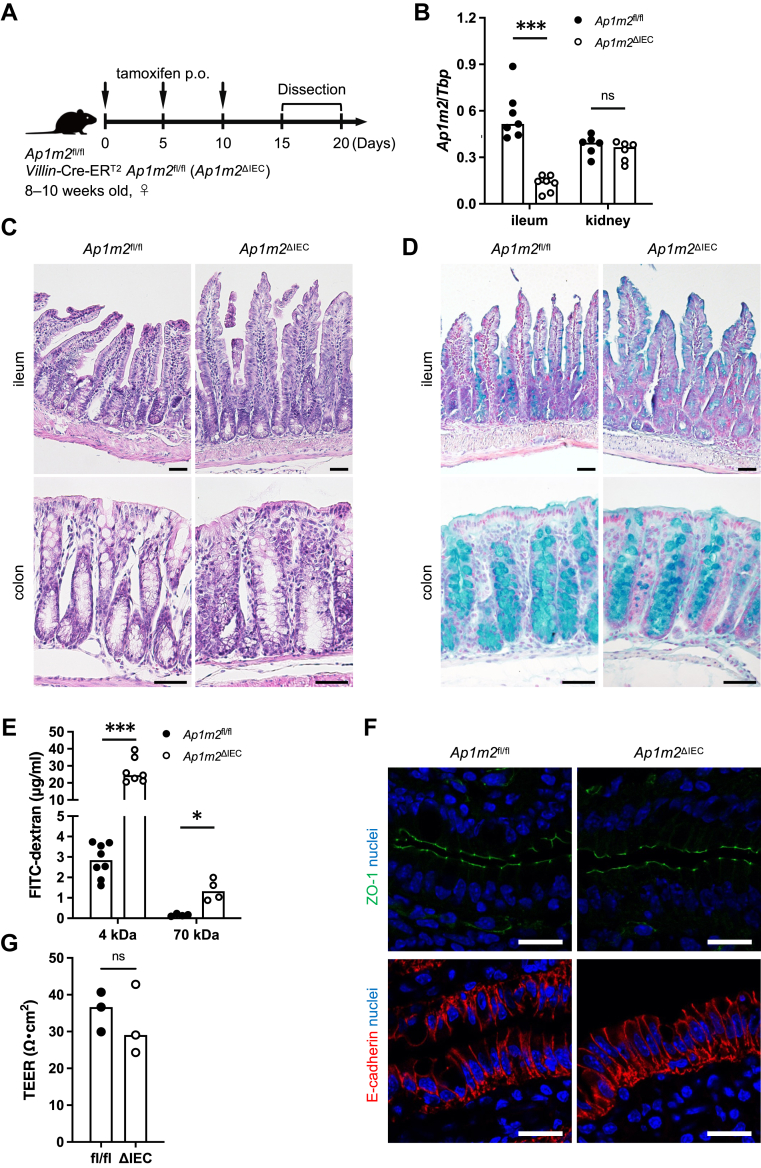
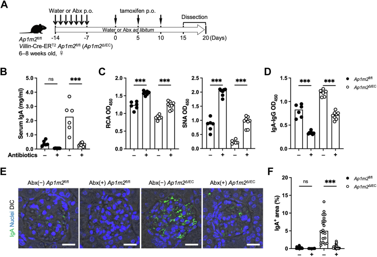
Methods: We generated IEC-specific Ap1m2-deficient (Ap1m2ΔIEC) mice with low intestinal barrier integrity as an LSG model and examined the systemic impact.

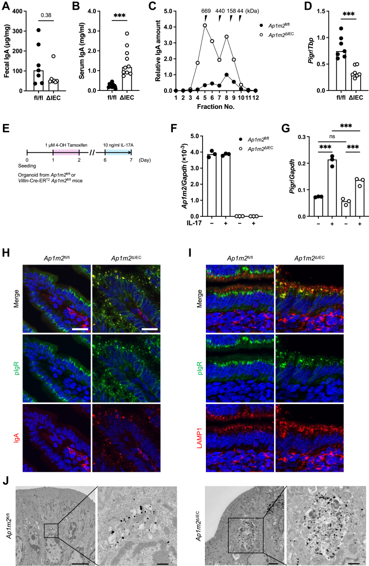
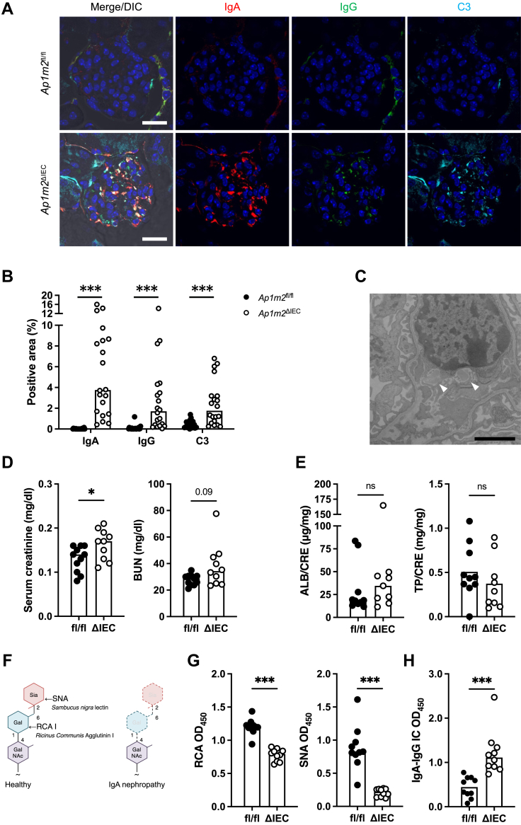
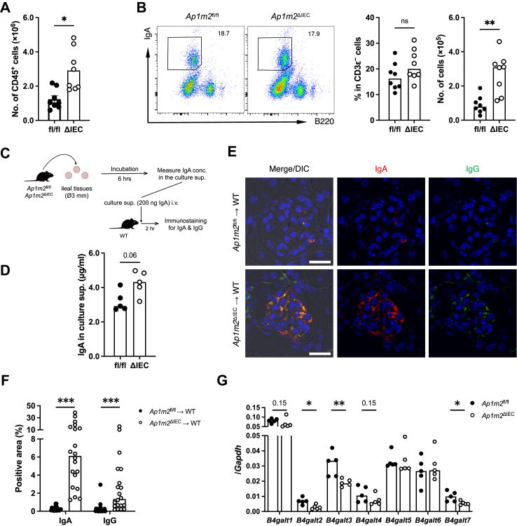
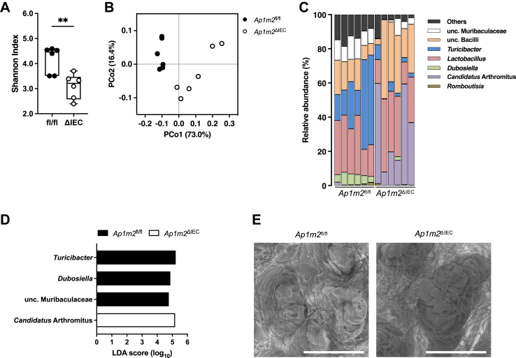
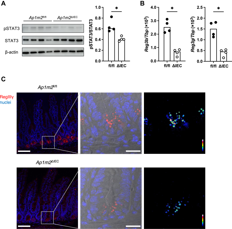
Findings: Ap1m2ΔIEC mice spontaneously developed IgA nephropathy (IgAN)-like features characterized by the deposition of IgA-IgG immune complexes and complement factors in the kidney glomeruli. Ap1m2 deficiency markedly enhanced aberrantly glycosylated IgA in the serum owing to downregulation and mis-sorting of polymeric immunoglobulin receptors in IECs. Furthermore, Ap1m2 deficiency caused intestinal dysbiosis by attenuating IL-22-STAT3 signaling. Intestinal dysbiosis contributed to the pathogenesis of IgAN because antibiotic treatment reduced aberrantly glycosylated IgA production and renal IgA deposition in Ap1m2ΔIEC mice.

Interpretation: IEC barrier dysfunction and subsequent dysbiosis by AP-1B deficiency provoke IgA deposition in the mouse kidney. Our findings provide experimental evidence of a pathological link between LGS and IgAN.

Funding: AMED, AMED-CREST, JSPS Grants-in-Aid for Scientific Research, JST CREST, Fuji Foundation for Protein Research, and Keio University Program for the Advancement of Next Generation Research Projects.

Keywords: Ap1m2; Epithelial barrier; IgA nephropathy; Intestinal microbiota; Leaky gut syndrome.


Conflict of interest statement

Declaration of interests The authors have declared that no conflict of interest exists.

Figures

Fig. 1
Fig. 1
The absence of AP-1B increases the intestinal permeability of dextran. (A) Experimental schedule of tamoxifen administration to Ap1m2fl/fl and Ap1m2ΔIEC mice for induction of Ap1m2 deficiency in the intestinal epithelial cells. Mice were orally administered 100 μl of 10 mg/ml tamoxifen diluted in sunflower seed oil every five days. Mice were used for the following experiments between days 15 and 20 after the initial administration. (B) Quantitative PCR analysis of Ap1m2 expression in the intestine and kidney from Ap1m2fl/fl and Ap1m2ΔIEC female mice after tamoxifen administration. Tbp was analyzed as a reference gene. n = 7 (ileum) or 6 (kidney). (C) and (D) Representative histological sections of (C) hematoxylin and eosin or (D) Alcian Blue-stained ileal and colonic tissues from Ap1m2fl/fl and Ap1m2ΔIEC female mice. Scale bars: 50 μm. (E) Intestinal permeability was assessed by the serum concentration of 4 kDa (n = 8 or 7) or 70 kDa (n = 4) FITC-dextran. (F) Immunofluorescence staining images of ZO-1 (green), E-cadherin (red), and nuclei (blue) in the ileum from Ap1m2fl/fl and Ap1m2ΔIEC female mice. Scale bars: 20 μm. (G) The trans-epithelial electrical resistance of isolated intestinal epithelium was measured with Ussing chamber. n = 3. Data represent the median. ns: not significant, ∗p < 0.05, ∗∗∗p < 0.001 (unpaired, two-tailed Mann–Whitney test).
Fig. 2
Fig. 2
AP-1B deficiency causes pIgR dysfunction and increases IgA in the blood. (A) and (B) IgA concentrations in feces in (A) and serum in (B) from Ap1m2fl/fl and Ap1m2ΔIEC female mice were measured by ELISA. n = 7 in (A) and n = 10 in (B) from two independent experiments. (C) Size exclusion chromatography fractionation profile of IgA in sera were pooled from Ap1m2fl/fl and Ap1m2ΔIEC female mice. The IgA levels in each fraction were measured by ELISA. (D) Quantitative PCR analysis of Pigr expression in the ileum from Ap1m2fl/fl and Ap1m2ΔIEC female mice. Tbp was analyzed as a reference gene. n = 7. (E) Experimental schedule for a treatment of intestinal organoids with IL-17. Organoids were generated from the ileum of Ap1m2fl/fl and Villin-Cre-ERT2Ap1m2fl/fl mice and treated with 1 μM 4-hydroxytamoxifen on day 1–2, followed 10 ng/ml recombinant IL-17A on day 6–7. (F) and (G) Quantitative PCR analysis of Ap1m2 (F) and Pigr (G) expression in Ap1m2fl/fl or Ap1m2ΔIEC organoids. Gapdh was analyzed as a reference gene. (H) and (I) Immunofluorescence images of pIgR (green), IgA or LAMP1 (red), and nuclei (blue) in the ileum from Ap1m2fl/fl and Ap1m2ΔIEC female mice. Scale bars: 20 μm. (J) Immuno-electron microscopic images of epithelial cells in the ileum from Ap1m2fl/fl and Ap1m2ΔIEC female mice for pIgR. Scale bars: 0.5 μm (left) or 1.0 μm (right). Data represent the median. ns: not significant, ∗∗∗p < 0.001 (unpaired, two-tailed Mann–Whitney test for comparison of two groups or two-way ANOVA with Bonferroni's multiple comparison test for comparison of multiple groups).
Fig. 3
Fig. 3
IgA–IgG immune complex is deposited in the glomeruli of Ap1m2ΔIECfemale mice. (A) Immunofluorescence images of IgA (red), IgG (green), complement C3 (cyan), and nuclei (blue) in the kidney from Ap1m2fl/fl and Ap1m2ΔIEC female mice. Scale bars: 20 μm. (B) Quantitative data of (A): the percentage of IgA+, IgG+, or C3+ area in a glomerulus area were quantified. n = 20, five glomeruli each from four different mice. (C) A representative transmission electron microscopic image of the mesangial cells in the kidney from Ap1m2ΔIEC female mice. Arrowheads indicate electron-dense deposits in the mesangial area. Scale bar: 2.0 μm. (D) Creatinine and blood urea nitrogen concentration in their sera of Ap1m2fl/fl (n = 11) and Ap1m2ΔIEC (n = 10) female mice from two independent experiments. (E) Urinary ratio of albumin and total protein to creatinine in Ap1m2fl/fl (n = 10) and Ap1m2ΔIEC (n = 9) female mice. (F) Illustration of lectin-binding assay. When IgA is completely glycosylated, galactose is added to N-acetylgalactosamine, followed by the addition of sialic acid. Therefore, IgA is recognized by Ricinus communis agglutinin I (RCA) or Sambucus nigra lectin (SNA). (G) Quantification of galactose or sialic acid in the serum IgA glycans by lectin-binding assay. 100 ng serum IgA was reacted with RCA or SNA, and the optical density (OD) was measured. n = 10 from two independent experiments. (H) Quantification of IgA-IgG immune complex (IC) in the serum. Serum IgA-IgG immune complex captured by anti-IgG antibody was detected with anti-IgA antibody. The bar graphs show optical density values at 450 nm. n = 10 from two independent experiments. Data represent the median. ns: not significant, ∗p < 0.05, ∗∗∗p < 0.001 (unpaired, two-tailed Mann–Whitney test).
Fig. 4
Fig. 4
Intestinal-derived IgA causes deposition in the kidney. (A) and (B) Flow cytometric analysis of leucocytes isolated from the ileal lamina propria of Ap1m2fl/fl and Ap1m2ΔIEC female mice. The total number of CD45+ cells (A); representative plots, the frequency and the number of IgA+ B cells (B). n = 8 from two independent experiments. (C) Experimental procedure: (D) Ileal tissues were sampled from Ap1m2fl/fl and Ap1m2ΔIEC female mice and were cultured for 6 h. Culture supernatant was collected, and IgA secreted into the supernatant was measured by ELISA. n = 5. and (E) The pooled culture supernatant of each group was adjusted to contain 200 ng IgA and injected intravenously into wild-type female mice. After 2 h, immunofluorescence analysis was performed. Images show IgA (red), IgG (green), and nuclei (blue) in the kidney from culture supernatant-treated wild-type female mice. Scale bars: 20 μm. (F) Quantitative data of (D): the percentage of IgA+ or IgG+ area in a glomerulus area. n = 20, five glomeruli each from four different mice. (G) Quantitative PCR analysis of B4galt1 to 7 expressions in B cells isolated from the ileal lamina propria of Ap1m2fl/fl and Ap1m2ΔIEC female mice. Gapdh was analyzed as a reference gene. n = 5. Data represent the median. ns: not significant, ∗p < 0.05, ∗∗p < 0.01, ∗∗∗p < 0.001 (unpaired, two-tailed Mann–Whitney test).
Fig. 5
Fig. 5
Ap1m2ΔIECmice exhibit dysbiosis in the gut microbiota. Microbiota profiles in the ileum from Ap1m2fl/fl and Ap1m2ΔIEC female mice. n = 6. (A) Species richness and evenness (Shannon index). Data represent the median, interquartile range, and minimum and maximum values. ∗∗p < 0.01 (unpaired, two-tailed Mann–Whitney test). (B) Principal coordinate analysis based on the weighted UniFrac analysis is shown. (C) Genus-level taxonomic distribution of individual mice. Values represent relative abundance (%). (D) Differentially abundant taxa at the genus level with an average relative abundance >0.3%. Linear discriminant analysis effect size (LEfSe) shows significantly different genera (LDA score >2). (E) Scanning electron microscopic image of ileal epithelium from Ap1m2fl/fl and Ap1m2ΔIEC female mice. Scale bars: 100 μm.
Fig. 6
Fig. 6
Gut microbiota contribute to the development of IgA nephropathy in Ap1m2ΔIECmice. (A) Experimental schedule of antibiotic treatment on Ap1m2fl/fl (n = 6 or 7) and Ap1m2ΔIEC (n = 6 or 8) female mice from two independent experiments. Antibiotics were administered in their drinking water ad libitum from the first two weeks before tamoxifen administration until dissection for experiments. During the first week, the antibiotic cocktail was additionally given orally once daily. (B) Serum IgA concentrations were measured by ELISA. (C) Serum IgA glycosylation was evaluated by lectin binding assay. (D) Serum IgA-IgG immune complex (IC) was quantified. (E) Immunofluorescence image of IgA (green) in kidneys. Scale bars: 50 μm. (F) Quantitative data of (D): the percentage of IgA+ area in a glomerulus area. n = 20, five glomeruli each from four different mice. Data represent the median. ns: not significant, ∗∗∗p < 0.001 (two-way ANOVA with Bonferroni's multiple comparison test).
Fig. 7
Fig. 7
Loss of AP-1B decreases IL-22-STAT3 signal, leading to decreased RegIIIγ expression in the intestine. (A) Western blot analysis of pSTAT3, STAT3, and β-actin in the intestinal epithelium from Ap1m2fl/fl and Ap1m2ΔIEC female mice. n = 4. (B) Quantitative PCR analysis of Reg3b and Reg3g in the intestine from ileal Ap1m2fl/fl and Ap1m2ΔIEC female mice. Tbp was analyzed as a reference gene. n = 4. (C) Immunofluorescence images of RegIIIγ (red) and nuclei (blue) in the intestine from Ap1m2fl/fl and Ap1m2ΔIEC female mice. Scale bars: 100 μm. Data represent the median. ∗p < 0.05 (unpaired, two-tailed Mann–Whitney test).

References

    1. Peterson L.W., Artis D. Intestinal epithelial cells: regulators of barrier function and immune homeostasis. Nat Rev Immunol. 2014;14:141–153. - PubMed
    1. Bevins C.L., Salzman N.H. Paneth cells, antimicrobial peptides and maintenance of intestinal homeostasis. Nat Rev Microbiol. 2011;9:356–368. - PubMed
    1. Johansson M.E.V., Hansson G.C. Immunological aspects of intestinal mucus and mucins. Nat Rev Immunol. 2016;16:639–649. - PMC - PubMed
    1. Rojas R., Apodaca G. Immunoglobulin transport across polarized epithelial cells. Nat Rev Mol Cell Biol. 2002;3:944–956. - PubMed
    1. Wei M., Shinkura R., Doi Y., Maruya M., Fagarasan S., Honjo T. Mice carrying a knock-in mutation of Aicda resulting in a defect in somatic hypermutation have impaired gut homeostasis and compromised mucosal defense. Nat Immunol. 2011;12:264–270. - PubMed

MeSH terms

Substances