. 2024 Aug 13;19(8):1061-1073.
doi: 10.1016/j.stemcr.2024.06.010. Epub 2024 Jul 25.

Skeletal muscle-on-a-chip in microgravity as a platform for regeneration modeling and drug screening

Affiliations

Skeletal muscle-on-a-chip in microgravity as a platform for regeneration modeling and drug screening

Soochi Kim et al. Stem Cell Reports. .

Abstract

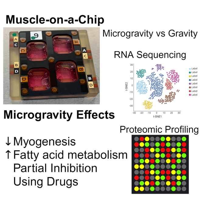
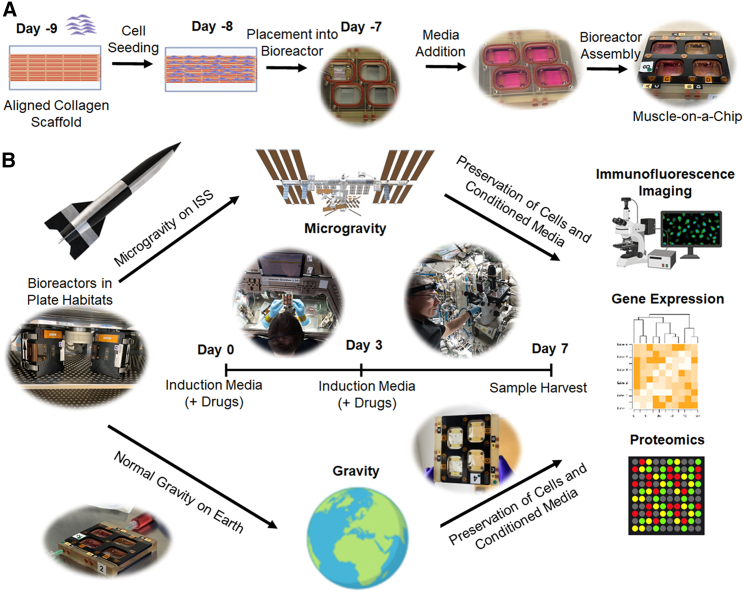
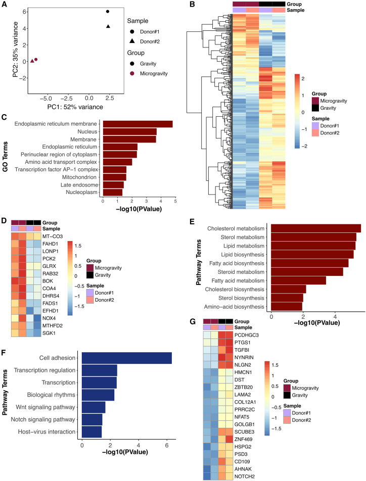
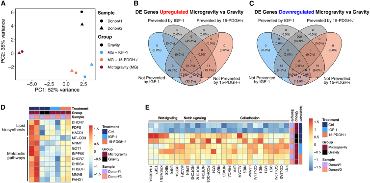
Microgravity has been shown to lead to both muscle atrophy and impaired muscle regeneration. The purpose was to study the efficacy of microgravity to model impaired muscle regeneration in an engineered muscle platform and then to demonstrate the feasibility of performing drug screening in this model. Engineered human muscle was launched to the International Space Station National Laboratory, where the effect of microgravity exposure for 7 days was examined by transcriptomics and proteomics approaches. Gene set enrichment analysis of engineered muscle cultured in microgravity, compared to normal gravity conditions, highlighted a metabolic shift toward lipid and fatty acid metabolism, along with increased apoptotic gene expression. The addition of pro-regenerative drugs, insulin-like growth factor-1 (IGF-1) and a 15-hydroxyprostaglandin dehydrogenase inhibitor (15-PGDH-i), partially inhibited the effects of microgravity. In summary, microgravity mimics aspects of impaired myogenesis, and the addition of these drugs could partially inhibit the effects induced by microgravity.

Keywords: aging; atrophy; microgravity; regeneration; skeletal muscle; tissue engineering; transcriptomics.


Conflict of interest statement

Declaration of interests The authors declare no competing interests.

Figures

None
Graphical abstract
Figure 1
Figure 1
Generation and characterization of biomimetic skeletal muscle-on-a-chip platform (A) Schematic depicting the fabrication of aligned collagen nanofibrils using shear-mediated extrusion of monomeric collagen into pH 7 buffer. (B) Gross image and schematic of anisotropic collagen strips deposited onto hydrophobic glass chips. (C) Scanning electron microscopy analysis shows highly organized anisotropic collagen fibrils. (D) Seeding of primary human skeletal muscle cells shows cell alignment along the direction of the collagen nanofibrils (denoted by arrow). (E) In induction media, the cells fuse to form parallel-aligned myotubes, as shown by myosin heavy-chain staining (green). Scale bars: (B) 5 mm, (C) 1 μm, (D–E) 100 μm.
Figure 2
Figure 2
Preparation of muscle-on-a-chip devices for microgravity studies and experimental overview (A) The overview of muscle-on-a-chip preparation consists of seeding human muscle cells onto immobilized aligned nanofibrillar collagen scaffolds followed by cell seeding. After one day, the cell-seeded scaffolds are placed into bioreactors and filled with growth media. The bioreactor is then secured with screws for transportation to the ISSNL. (B) The experimental overview consists of incubating the bioreactor during transit to the ISSNL in plate habitats that provide temperature maintenance during transport to the ISSNL or in normal gravity conditions on Earth. Upon docking at the ISSNL, microgravity experiments began (day 0) with media change into an induction media that supports myotube formation. In some experiments, drugs were added to the media on day 0 and again on day 3. On day 7, the conditioned media were extracted, and the samples were stabilized in RNAlater or fixed in paraformaldehyde for analysis on by RNA sequencing, proteomics analysis, or myotube formation.
Figure 3
Figure 3
Transcriptional differences between microgravity and gravity control biomimetic skeletal muscle (A) Principal-component analysis of the transcriptome of biomimetic skeletal muscle samples after 7 days in microgravity or control conditions (gravity). (B) Heatmap shows all differentially expressed genes after 7 days in microgravity, compared to gravity (FDR 10%). (C) Gene Ontology (GO) enrichment terms of differentially upregulated genes in microgravity (p < 0.05). (D) Heatmap shows the expression of genes found in the mitochondrion GO enrichment term. (E) Pathway enrichment terms of differentially upregulated genes in microgravity (p < 0.05). (F) Pathway enrichment terms of differentially downregulated genes in microgravity (p < 0.05). (G) Heatmap shows expression of top 20 differentially downregulated genes in microgravity (n = 2 donors per group).
Figure 4
Figure 4
Transcriptional similarities between clinical sarcopenia muscle and engineered skeletal muscle in microgravity (A) Principal-component analysis of the transcriptome in healthy vs. sarcopenia clinical samples from both sexes. (B) Volcano plot shows differentially expressed genes in sarcopenia, compared to normal conditions (p < 0.05, log2 FC > 1.0). Genes involved in DNA damage response, extracellular matrix organization, VEGFA-VEGFR2 signaling pathway, and Wnt signaling are labeled. Genes showing statistically significant expression (FDR 25%) are indicated in red. (C) Heatmap shows the genes involved in Wnt signaling, rhabdomyosarcoma, DNA damage response, extracellular matrix organization, and VEGFA-VEGFR2 signaling. (D–F) Enrichment plots for custom gene set enrichment analysis (GSEA). The plots report positive enrichment of Old_MuSC_UP gene set (D) and negative enrichment of Sarcopenia_UP and Sarcopenia-DOWN gene sets (E–F) in microgravity, compared to gravity samples. The enrichment plot shows the gene set names (top) and the running enrichment score. The red curve indicates positive enrichment, whereas the blue curve indicates negative enrichment scores. Black bars indicate the positions of the gene set on the rank-ordered list in GSEA, and the signal-to-noise metrics are shown at the bottom.
Figure 5
Figure 5
IGF-1 and 15-PDGFH inhibitor treatment can partially prevent microgravity effect (A) Principal-component analysis of transcriptome in 7 days in microgravity (MG) or gravity control conditions in the presence of drug treatment (IGF-1 or 15-PDGH-i) in biomimetic skeletal muscle samples. (B) The 4-way Venn diagram shows the number of differentially expressed genes that were upregulated in microgravity, compared to gravity conditions, or whose differential expression could be either prevented by IGF-1 or 15-PDGH-i treatment (gray) or not prevented by drug treatment (IGF-1 in blue and 15-PDGH-i in orange). (C) 4-way Venn diagram showing the number of differentially expressed genes downregulated in microgravity, compared to gravity conditions, or whose differential expression could be either prevented by IGF-1 or 15-PDGH-i treatment (gray) or not prevented by drug treatment (IGF-1 in blue and 15-PDGH-i in orange). (D) Heatmap showing genes implicated in lipid biosynthesis, metabolic pathways, and adipogenesis. (E) Heatmap showing genes implicated in cell adhesion, Notch signaling, and Wnt signaling (n = 2 donors per group).

References

    1. Alcazar C.A., Hu C., Rando T.A., Huang N.F., Nakayama K.H. Transplantation of insulin-like growth factor-1 laden scaffolds combined with exercise promotes neuroregeneration and angiogenesis in a preclinical muscle injury model. Biomater. Sci. 2020;8:5376–5389. doi: 10.1039/d0bm00990c. - DOI - PMC - PubMed
    1. Andres-Mateos E., Brinkmeier H., Burks T.N., Mejias R., Files D.C., Steinberger M., Soleimani A., Marx R., Simmers J.L., Lin B., et al. Activation of serum/glucocorticoid-induced kinase 1 (SGK1) is important to maintain skeletal muscle homeostasis and prevent atrophy. EMBO Mol. Med. 2013;5:80–91. doi: 10.1002/emmm.201201443. - DOI - PMC - PubMed
    1. Arthur S.T., Cooley I.D. The effect of physiological stimuli on sarcopenia; impact of Notch and Wnt signaling on impaired aged skeletal muscle repair. Int. J. Biol. Sci. 2012;8:731–760. doi: 10.7150/ijbs.4262. - DOI - PMC - PubMed
    1. Ascenzi F., Barberi L., Dobrowolny G., Villa Nova Bacurau A., Nicoletti C., Rizzuto E., Rosenthal N., Scicchitano B.M., Musaro A. Effects of IGF-1 isoforms on muscle growth and sarcopenia. Aging Cell. 2019;18:e12954. doi: 10.1111/acel.12954. - DOI - PMC - PubMed
    1. Bjornson C.R., Cheung T.H., Liu L., Tripathi P.V., Steeper K.M., Rando T.A. Notch signaling is necessary to maintain quiescence in adult muscle stem cells. Stem Cell. 2012;30:232–242. doi: 10.1002/stem.773. - DOI - PMC - PubMed

Publication types

MeSH terms

Substances