. 2024 Sep;300(9):107601.
doi: 10.1016/j.jbc.2024.107601. Epub 2024 Jul 24.

CYYR1 promotes the degradation of the E3 ubiquitin ligase WWP1 and is associated with favorable prognosis in breast cancer

Affiliations

CYYR1 promotes the degradation of the E3 ubiquitin ligase WWP1 and is associated with favorable prognosis in breast cancer

Tiphaine Perron et al. J Biol Chem. 2024 Sep.

Abstract

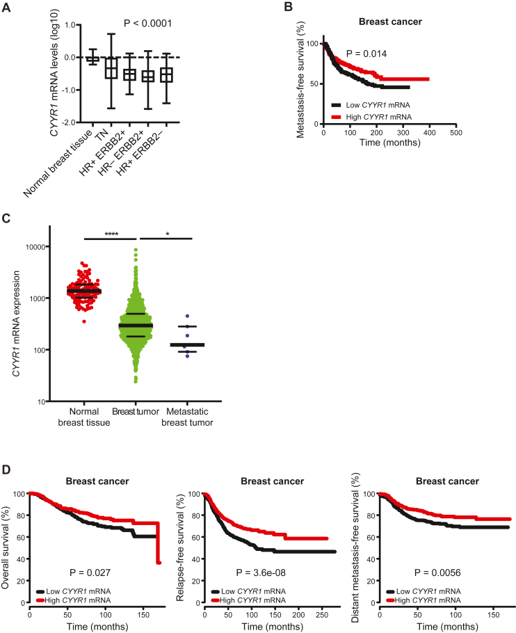
Ubiquitination plays a crucial role in cellular homeostasis by regulating the degradation, localization, and activity of proteins, ensuring proper cell function and balance. Among E3 ubiquitin ligases, WW domain-containing protein 1 (WWP1) is implicated in cell proliferation, survival, and apoptosis. Notably WWP1 is frequently amplified in breast cancer and associated with poor prognosis. Here, we identify the protein cysteine and tyrosine-rich protein 1 (CYYR1) that had previously no assigned function, as a regulator of WWP1 activity and stability. We show that CYYR1 binds to the WW domains of the E3 ubiquitin ligase WWP1 through its PPxY motifs. This interaction triggers K63-linked autoubiquitination and subsequent degradation of WWP1. We furthermore demonstrate that CYYR1 localizes to late endosomal vesicles and directs polyubiquitinated WWP1 toward lysosomal degradation through binding to ANKyrin repeat domain-containing protein 13 A (ANKRD13A). Moreover, we found that CYYR1 expression attenuates breast cancer cell growth in anchorage-dependent and independent colony formation assays in a PPxY-dependent manner. Finally, we highlight that CYYR1 expression is significantly decreased in breast cancer and is associated with beneficial clinical outcome. Taken together our study suggests tumor suppressor properties for CYYR1 through regulation of WWP1 autoubiquitination and lysosomal degradation.

Keywords: CYYR1; E3 ubiquitin ligase; WWP1; breast cancer; ubiquitination.


Conflict of interest statement

Conflict of interest The authors declare that they have no conflicts of interest with the contents of this article.

Figures

Figure 1
Figure 1
CYYR1 interacts with WWP1 E3 ubiquitin ligases.A, association of CYYR1 with NEDD4 family E3s. HEK293 cells were transfected with HA-CYYR1 either alone or with Flag-tagged catalytically inactive E3s as indicated. Cell lysates immunoprecipitated with anti-Flag antibody and total cell lysates (TCLs) were analyzed by Western blotting with the indicated antibodies. A schematic representation of the NEED4 family E3s showing the C2, WW, and HECT domain. B, association between endogenous CYYR1 and WWP1. MDA-MB-468 total cell lysate immunoprecipitated or not with anti-CYYR1 or IgG antibody were analyzed by Western blotting as indicated. C, proximity of CYYR1 and WWP1. Proximity ligation assay (PLA) experiments were performed in MDA-MB-468 cells in presence of the indicated antibodies. Antibodies alone conditions are used as a negative control. Dapi staining is shown in the lower panel. The bar represents 10 μm. Statistical analysis of the number of dots/cell was performed on 50 cells of one representative experiment using one-way ANOVA followed by Sidak’s test. D, schematic representation of the WWP1 constructs showing the C2, WW, and HECT domains. Lower panel: WWP1 binding domain to CYYR1. HEK293 cells were transfected with HA-CYYR1 either alone or with full length or domains of Flag-WWP1, as indicated. Cell lysates immunoprecipitated with anti-Flag antibody were analyzed as in A. E, schematic representation of the CYYR1 constructs. PS (peptide signal), CYS-Rich (cystein-rich domain), TM (transmembrane domain) and PPxY motifs 1 to 3 are represented. Lower panel: CYYR1 binding domain to WWP1. HEK293 cells were transfected with Flag-WWP1-CA either alone or with HA-CYYR1-ΔPPxY mutants as indicated. Cell lysates immunoprecipitated with anti-HA antibody were analyzed as described. CA, catalytically inactive; CYYR1, cysteine and tyrosine-rich protein 1; HA, hemagglutinin; HECT, homologous to E6AP C terminus; NEDD4, neural precursor cell-expressed developmentally downregulated 4; WWP1, WW domain-containing protein 1.
Figure 2
Figure 2
CYYR1 is ubiquitinated by WWP1 and WWP2 at lysine K154.A and B, CYYR1 is ubiquitinated by WWP1 and WWP2 at lysine K154. HEK293 cells were transfected with HA-CYYR1-WT or HA-CYYR1-K154R either alone or with His-Ubiquitin (His-Ub) and Flag-tagged WWP1 or WWP2 (WT) or their catalytically inactive mutants (CA) and treated with MG132 for 4 h before lysis. Cell lysates were pulled-down with the ubiquitin pan Selector affinity resin and analyzed by Western blotting with the indicated antibodies. Western blotting on TCL is shown as a transfection control. C, CYYR1-K154R binds to WWP1 and WWP2. Cell lysates from HEK293 cells transfected with GFP-CYYR1 or GFP-CYYR1-K154R were immunoprecipitated with the GFP-trap affinity resin and analyzed by Western blotting as indicated. D, MDA-MB-468 cells were transfected with a nontargeted siRNA control (siNT) or two independent siRNA (#1 and #2) targeting WWP1 or WWP2. Seventy-two hours posttransfection, cell lysates were analyzed by Western blotting. Quantifications of the CYYR1 intensity relative to GAPDH intensity in each condition normalized to the siNT control condition and p-values were calculated using one-way ANOVA followed by Dunnett’s test (n = 3). ∗ on anti-WWP2 Western blot indicates nonspecific band. CYYR1, cysteine and tyrosine-rich protein 1; HA, hemagglutinin; TCL, total cell lysate; WWP1/2, WW domain-containing protein 1/2.
Figure 3
Figure 3
CYYR1 regulates WWP1 autoubiquitination and protein level.A, CYYR1 increases WWP1 autoubiquitination. HEK293 cells transfected with His-Ub and Flag-WWP1-WT or Flag-WWP1-CA either alone or with different HA-CYYR1 constructs as indicated. Protein cell lysates were pulled-down with the ubiquitin pan Selector affinity resin and analyzed by Western blotting. B, CYYR1 decreases WWP1 protein level. Cell lysates form HEK293 cells transfected with Flag-WWP1-WT or Flag-WWP1-CA either alone or with different constructs for HA-CYYR1 were analyzed by Western blotting. C, CYYR1 depletion increases WWP1 protein level. Cell lysates from MDA-MB-468 cells transfected with a nontargeting siRNA control (siNT) or two independent siRNA (#1 or #2) targeting CYYR1 were analyzed by Western blotting using the indicated antibodies. Quantifications of the WWP1 intensity relative to GAPDH in each condition were normalized to the siNT control condition, and p-values were calculated with a one-way ANOVA followed by Dunnett’s test (n = 5). D, CYYR1 decreases endogenous WWP1 protein level. MDA-MB-231 cells expressing doxycycline (Dox)-inducible CYYR1-WT or CYYR1-ΔPPxY (TO-CYYR1-WT or TO-CYYR1-ΔPPxY clones #1 and #2) were treated with Dox for 24 h before analysis of the cell lysates by Western blotting. Low and high exposure of the anti-CYYR1 Western blot are shown to highlight CYYR1 expression leakiness in absence of Dox. Quantifications of the WWP1 intensity relative to GAPDH in each condition were normalized to the Ctrl condition and p-values were calculated with one-way ANOVA followed by Dunnett’s test (n = 3). CYYR1, cysteine and tyrosine-rich protein 1; Dox, doxycycline; WWP1, WW domain-containing protein 1.
Figure 4
Figure 4
CYYR1 induces lysosome-dependent degradation of WWP1.A, CYYR1 induces K63-linked polyubiquitination of WWP1. HEK293 cells were transfected with Flag-WWP1-WT either alone or with different constructs for HA-CYYR1 as indicated. Cell lysates were pulled-down with the ubiquitin pan, the ubiquitin K63 or the ubiquitin K48 Selector affinity resins and analyzed by Western blotting using the indicated antibodies. B, CYYR1 induces polyubiquitination of endogenous WWP1. MDA-MB-231 TO-Ctrl or TO-CYYR1-WT clones #1 and #2 were treated with Dox 10 ng/ml and with bafilomycin A1 (BAF) 100 nM and chloroquine (CQ) 50 μM or DMSO for 24 h before lysis. Cell lysates were pulled-down with the ubiquitin pan Selector affinity resins and analyzed by Western blotting using the indicated antibodies. C, CYYR1 colocalizes with GFP-Rab7A. HeLa cells transfected with untagged CYYR1 and GFP-Rab7A (green) were stained by immunofluorescence with anti-CYYR1 antibody (blue). Channel intensity plots along a 10 μm line is shown. D, WWP1 colocalizes with CYYR1 at GFP-Rab7A-tagged late endosomes. HeLa cells transfected with Flag-WWP1-CA and GFP-Rab7A (green) in presence or in absence of untagged CYYR1-WT or CYYR1-ΔPPxY, were subjected to immunofluorescence with anti-CYYR1 (blue) and anti-Flag (red). E, CYYR1 targets WWP1 to GFP-Rab7A-tagged late endosomes. The % of colocalization of Flag-WWP1-CA with GFP-Rab7A was measured in each condition on 20 cells in three independent experiments. This percentage was determined by measuring the intensity of anti-Flag staining within the perinuclear GFP-Rab7-stained area relative to the overall anti-Flag staining intensity of each cell. Statistical analysis was performed with one-way ANOVA followed by Sidak’s test. The scale bar in each image represents 10 μm. The scale bar in the enlarged image represents 5 μm. CYYR1, cysteine and tyrosine-rich protein 1; DMSO, dimethyl sulfoxide; Dox, doxycycline.
Figure 5
Figure 5
ANKRD13A is involved in CYYR1-induced WWP1 degradation.A, CYYR1-interactome. Cell lysates from HEK293 cells transfected with GFP or GFP-CYYR1 were purified on GFP-Trap affinity resin and analyzed by quantitative label-free mass spectrometry. Volcano plot showing differentially expressed proteins significantly enriched in GFP-CYYR1 compared to GFP (green line correspond to fold change ≥ 2, and red line to p-value ≤ 0.05, n = 5) that display at least three peptides in each of the five replicate experiments. Proteins that display no common peptides in the GFP condition (infinite ratio) are represented on the left of the graph with their distribution based on the number of peptides identified/100aa. B, CYYR1 interacts with ANKRD13A. Cell lysates from HEK293 cells transfected with GFP or GFP-CYYR1 were purified with GFP-Trap affinity resin and analyzed by Western blotting with the indicated antibodies. C, WWP1 interacts with ANKRD13A in the presence of CYYR1. HEK293 cells were transfected with Flag-WWP1 or Flag-WWP1-CA and HA-CYYR1-WT or HA-CYYR1-ΔPPxY with or without Flag-ANKRD13A. Cell lysates were immunoprecipitated with anti-ANKRD13A antibody and immunoblotted with the indicated antibodies. D, schematic representation of the ANKRD13A constructs. AR and UIM domains are shown. Lower panel: ANKRD13A binds WWP1 through its UIM domains. HEK293 cells were transfected with HA-CYYR1, Flag-ANKRD13A-WT, or deletion mutants and GFP or GFP-WWP1-WT as indicated. Cell lysates were immunoprecipitated with the GFP-trap affinity resin and immunoblotted with the corresponding antibodies. E, schematic representation of the ANKRD13A, B, C, and D. Lower panel: WWP1 binds all members of the ANKRD13 family except ANKRD13C. Cell lysates from HEK293 cells were transfected with HA-CYYR1 and Flag-tagged ANKRD13 family proteins, and GFP or GFP-WWP1-WT were immunoprecipitated with the GFP-trap affinity resin and analyzed by Western blotting with the indicated antibodies. F, ANKRD13A depletion attenuates the degradation of WWP1 induced by CYYR1. MDA-MB-231 TO-CYYR1-WT clone #1 were transfected with control siRNA (siNT) or two independent siRNA targeting ANKRD13A. Seventy-two hours post transfection, cells were treated with 500 pg/ml Dox for 24 h before lysis and Western blotting analysis with the indicated antibodies. Quantifications of the WWP1 intensity relative to GAPDH in each + Dox condition were normalized to the -Dox condition and p-values were calculated by performing a paired t test, ∗p < 0.05 (n = 3). ANKRD13A, ANKyrin repeat domain-containing protein 13 A; AR, ankyrin-repeat; CYYR1, cysteine and tyrosine-rich protein 1; Dox, doxycycline; UIM, ubiquitin-interacting motif; WWP1, WW domain-containing protein 1.
Figure 6
Figure 6
CYYR1 expression limits anchorage-dependent and independent colony formation of MDA-MB-231 breast cancer cells.A, MDA-MB-231 cells were transfected with pMEP4-CYYR1-WT or pMEP4-CYYR1-ΔPPxY before selection with hygromycin B. Colonies were stained with crystal violet and counted. Statistical analysis was performed with one-way ANOVA followed by Dunnett’s test (Ctrl versus CYYR1-WT, n = 4; Ctrl versus CYYR1-ΔPPxY, n = 3). B, MDA-MB-231 TO-Ctrl, TO-CYYR1-WT or -ΔPPxY clones were plated for soft agar growth assay in presence or absence of doxycycline. Colonies were stained with Thiazolyl Blue Tetrazolium Bromide coloration and counted. The % of colonies obtained in each condition relative to the TO-Ctrl condition are represented (Ctrl versus CYYR1-WT, n = 6 independent experiments; Ctrl versus CYYR1-ΔPPxY, n = 3). Statistical analysis was performed with one-way ANOVA followed by Dunnett’s test. CYYR1, cysteine and tyrosine-rich protein 1.
Figure 7
Figure 7
CYYR1 expression is decreased in breast cancer and associated with beneficial clinical outcome.A, analysis of the mRNA level of CYYR1 by RT-qPCR on normal breast tissue and four different human breast tumor subtypes, triple-negative breast cancer (TN; HR-ERBB2-), hormone receptor positive/HER2 positive (HR + ERBB2+), hormone receptor negative/HER2 positive (HR-ERBB2+) and hormone receptor positive/HER2 negative (HR + ERBB2-) in a cohort of 505 breast cancer patients. p-value was calculated using the Kruskal-Wallis H test. B, Kaplan-Meier analysis of metastasis-free survival rates of patients with tumors expressing high (red line) versus low (black line) levels of CYYR1 mRNA expression in the same cohort of 505 breast cancer patients. C, mRNA level of CYYR1 from the KM plotter database in normal breast tissues, breast tumor tissues and metastatic breast tumor tissues. D, Kaplan-Meier analyses of overall survival, relapse-free survival or distant metastasis-free survival according to CYYR1 mRNA expression in KM plotter database of breast cancer. CYYR1, cysteine and tyrosine-rich protein 1; RT-qPCR, quantitative reverse transcription polymerase chain reaction.
Figure 8
Figure 8
Model for lysosome degradation of WWP1 mediated by CYYR1. CYYR1 localizes at late endosomes where it recruits WWP1 in a PPxY/WW dependent manner. This interaction triggers K63-linked auto-ubiquitination of WWP1 that allows subsequent binding of ANKRD13A in a Ubiquitin/UIM dependent manner. These interactions targets WWP1 toward lysosomal degradation resulting in an overall decrease of WWP1 protein level. ANKRD13A, ANKyrin repeat domain-containing protein 13 A; CYYR1, cysteine and tyrosine-rich protein 1; UIM, ubiquitin-interacting motif; WWP1, WW domain-containing protein 1.

Similar articles

Cited by

References

    1. Swatek K.N., Komander D. Ubiquitin modifications. Cell Res. 2016;26:399–422. - PMC - PubMed
    1. Clague M.J., Urbé S. Ubiquitin: same molecule, different degradation pathways. Cell. 2010;143:682–685. - PubMed
    1. Buetow L., Huang D.T. Structural insights into the catalysis and regulation of E3 ubiquitin ligases. Nat. Rev. Mol. Cell Biol. 2016;17:626–642. - PMC - PubMed
    1. Wang Y., Argiles-Castillo D., Kane E.I., Zhou A., Spratt D.E. HECT E3 ubiquitin ligases - emerging insights into their biological roles and disease relevance. J. Cell Sci. 2020;133 - PMC - PubMed
    1. Ingham R.J., Gish G., Pawson T. The Nedd4 family of E3 ubiquitin ligases: functional diversity within a common modular architecture. Oncogene. 2004;23:1972–1984. - PubMed

Substances