. 2024 Sep 1:185:190-202.
doi: 10.1016/j.actbio.2024.07.014. Epub 2024 Jul 24.

Binary fabrication of decellularized lung extracellular matrix hybridgels for in vitro chronic obstructive pulmonary disease modeling

Affiliations

Binary fabrication of decellularized lung extracellular matrix hybridgels for in vitro chronic obstructive pulmonary disease modeling

Leigh-Ann M Antczak et al. Acta Biomater. .

Abstract

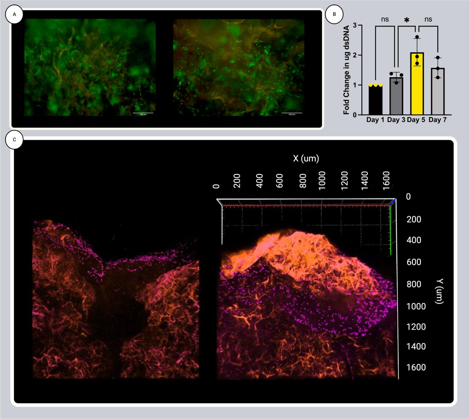
Limited treatments and a lack of appropriate animal models have spurred the study of scaffolds to mimic lung disease in vitro. Decellularized human lung and its application in extracellular matrix (ECM) hydrogels has advanced the development of these lung ECM models. Controlling the biochemical and mechanical properties of decellularized ECM hydrogels continues to be of interest due to inherent discrepancies of hydrogels when compared to their source tissue. To optimize the physiologic relevance of ECM hydrogel lung models without sacrificing the native composition we engineered a binary fabrication system to produce a Hybridgel composed of an ECM hydrogel reinforced with an ECM cryogel. Further, we compared the effect of ECM-altering disease on the properties of the gels using elastin poor Chronic Obstructive Pulmonary Disease (COPD) vs non-diseased (ND) human lung source tissue. Nanoindentation confirmed the significant loss of elasticity in hydrogels compared to that of ND human lung and further demonstrated the recovery of elastic moduli in ECM cryogels and Hybridgels. These findings were supported by similar observations in diseased tissue and gels. Successful cell encapsulation, distribution, cytotoxicity, and infiltration were observed and characterized via confocal microscopy. Cells were uniformly distributed throughout the Hybridgel and capable of survival for 7 days. Cell-laden ECM hybridgels were found to have elasticity similar to that of ND human lung. Compositional investigation into diseased and ND gels indicated the conservation of disease-specific elastin to collagen ratios. In brief, we have engineered a composited ECM hybridgel for the 3D study of cell-matrix interactions of varying lung disease states that optimizes the application of decellularized lung ECM materials to more closely mimic the human lung while conserving the compositional bioactivity of the native ECM. STATEMENT OF SIGNIFICANCE: The lack of an appropriate disease model for the study of chronic lung diseases continues to severely inhibit the advancement of treatments and preventions of these otherwise fatal illnesses due to the inability to recapture the biocomplexity of pathologic cell-ECM interactions. Engineering biomaterials that utilize decellularized lungs offers an opportunity to deconstruct, understand, and rebuild models that highlight and investigate how disease specific characteristics of the extracellular environment are involved in driving disease progression. We have advanced this space by designing a binary fabrication system for a ECM Hybridgel that retains properties from its source material required to observe native matrix interactions. This design simulates a 3D lung environment that is both mechanically elastic and compositionally relevant when derived from non-diseased tissue and pathologically diminished both mechanically and compositionally when derived from COPD tissue. Here we describe the ECM hybridgel as a model for the study of cell-ECM interactions involved in COPD.

Keywords: 3D disease modeling; Chronic obstructive pulmonary disease; Extracellular matrix; Lung disease; Tissue engineering.


Conflict of interest statement

Declaration of competing interests The authors declare the following financial interests/personal relationships which may be considered as potential competing interests: Rebecca L. Heie reports financial support was provided by National Institutes of Health. Dr. Heise has no other competing financial interests or personal relationships to disclose. Authors Antczak, Hendrick, and Moore state that they have no known competing financial interests or personal relationships that could have appeared to influence the work reported in this paper.

Figures

Figure 1.
Figure 1.
Matrix Composition. Quantification of major ECM components: insoluble collagen (A), soluble collagen (B), GAGs (C), and elastin (D), showing the relative reduction caused by the decellularization of human lung tissue. Data was normalized to the starting weight of dry lung tissue for the Intact Human Lung group and dry decellularized ECM working material for the ECM Source Material Group. Data is presented as mean +/− st.dev., n = 3 per group p < 0.05 as measured by t test. (E) Key matrix components presented as parts of a whole to describe the fractional relationship of the proteins to one another in intact human lung (pink) and decellularized ECM working material (grey).
Figure 2.
Figure 2.
Pore and Fiber Analysis of ND Human Lung and ECM Gels. SEM images comparing the structure of intact human lung tissue to human lung ECM cryogels prepared with 5, 10, and 20% concentrations of ECM working material (A) and the quantitative analysis of the effect of these concentrations on pore size(B). SEM images comparing the structure of an ECM hydrogel, ECM cryogel, and ECM hybridgel at 300, 100 and 100X magnification respectively (C). Pore size and fiber diameter analysis comparing ECM hydrogels, ECM cryogels, and intact human lung (D) and (E). **** p < 0.0001, *** p < .001, and * p < .05 for comparison of all groups via one-way ANOVA. Data is presented as mean +/− standard deviation (std. dev.) n = 3 for all groups.
Figure 3.
Figure 3.
Nanoindentation Mechanics. Representative force/strain curves of the spherical nanoindentation of ND human lungs, ECM hydrogels, cryogels, and hybridgels (A). Fold change in measured elastic moduli of ECM hydrogels, cryogels and hybridgels when compared to that of ND human lung as determined by the Hertz model (B). Fold change in measured hardness of ECM hydrogels, cryogels and hybridgels when compared to that of ND human lung as determined by martens hardness analysis (C). Data is presented as mean +/− std. dev. n = 3 per group. *p < 0.05, **p < 0.01, ***p < 0.001, ****p < 0.0001 for comparison of all groups via one-way ANOVA.
Figure 4.
Figure 4.
Encapsulation of hLFs. Confocal imaging of cellular distribution (A) and (B) at 500 μm depth of 1 mm ECM hybridgels. Cells stained for actin (red) while encapsulated in hydrogel (not visible) supported by the ECM cryogel that auto-fluoresces (blue) as described in schematic (C). 50 μm z-stack confocal experiment of horizontal hybridgel cross sections with encapsulated hLFs (D).
Figure 5.
Figure 5.
Proliferation of hLFs. Live/Dead assay fluorescent images (scale bar = 200 μm) of hybridgel cross sections after 3 and 5 days of culture (A). dsDNA picogreen assay of double stranded DNA isolated from hLFs encapsulated and cultured in hybridgels for 1, 3, 5, and 7 days (B). Confocal 1600 μm z-stack images of vertical ECM hybridgel cross sections with hLFs seeded directly on top. Cells were cultured for 1 (left) and 5 (right) days (C). Data is presented as mean +/− std. dev. n = 3 per group. *p < 0.05 for comparison of all groups via one-way ANOVA.
Figure 6.
Figure 6.
Cell Encapsulation’s Influence on Mechanics. Fold change in measured elastic moduli of cell-laden ECM hybridgels when compared to that of ND human lung as determined by the Hertz model (B). Fold change in measured hardness of cell laden ECM hybridgels when compared to that of ND human lung as determined by Martens Hardness analysis (C). Data is presented as mean +/− std. dev. n = 27 individual measurements per group, 3 gels per group with 9 measurements per gel.
Figure 7.
Figure 7.
Characterization of COPD ECM Hybridgels. Quantitative analysis of pore size and fiber formation of COPD ECM hydrogels and cryogels compared to that of ND ECM hydrogels and cryogels (A). SEM images comparing the structure of a COPD ECM hydrogel, cryogel (B) and hybridgel (C), at 300, 100 and 100X magnification respectively, to that of their respective ND gels. Quantification of major ECM components: insoluble collagen, soluble collagen, GAGs, and elastin in COPD ECM working material compared to that of ND ECM working material (D). Ratios of ND (grey) and COPD (yellow) intact human lung (dark) versus ECM working material (light) elastin to their respective groups of collagen (black) (E). Data is presented as mean +/− std. dev. n = 3 per group. *p < 0.05, **p < 0.01, ***p < 0.001, ****p < 0.0001 for comparison of all groups via two-way ANOVA (A), and t test or one-way ANOVA (D)
Figure 8.
Figure 8.
Nanoindentation Mechanics of Diseased Materials. Representative force/strain curves of the spherical nanoindentation of COPD ECM hydrogels, cryogels, and hybridgels compared to that of COPD lung. (A) Elastic moduli as determined by the Hertz model of COPD diseased human lung and ECM hydrogels, cryogels and hybridgels force/strain curves when compared to that of their respective ND groups (B). Hardness as determined by marten hardness analysis of COPD diseased human lung and ECM hydrogels, cryogels and hybridgels force/strain curves when compared to that of their respective ND groups (C). Data is presented as mean +/− std. dev. n = 3 per group. *p < 0.05, **p < 0.01, ***p < 0.001, ****p < 0.0001 for comparison of all groups via two-way ANOVA.

References

    1. Ahmad FB, Cisewski JA, Xu J, and Anderson RN, “Provisional Mortality Data — United States, 2022,” MMWR. Morb. Mortal. Wkly. Rep, vol. 72, no. 18, pp. 488–492, May 2023, doi: 10.15585/MMWR.MM7218A3. - DOI - PMC - PubMed
    1. Chaudhuri O, Cooper-White J, Janmey PA, Mooney DJ, and Shenoy VB, “Effects of extracellular matrix viscoelasticity on cellular behaviour,” Nat. 2020 5847822, vol. 584, no. 7822, pp. 535–546, Aug. 2020, doi: 10.1038/s41586-020-2612-2. - DOI - PMC - PubMed
    1. White ES, “Lung extracellular matrix and fibroblast function,” Ann. Am. Thorac. Soc, vol. 12, pp. S30–S33, Mar. 2015, doi: 10.1513/ANNALSATS.201406-240MG/SUPPL_FILE/DISCLOSURES.PDF. - DOI - PMC - PubMed
    1. Grolma JM, Weinand P, and Moone DJ, “Extracellular matrix plasticity as a driver of cell spreading,” Proc. Natl. Acad. Sci. U. S. A, vol. 117, no. 42, pp. 25999–26007, Oct. 2020, doi: 10.1073/PNAS.2008801117/SUPPL_FILE/PNAS.2008801117.SM02.MP4. - DOI - PMC - PubMed
    1. Burgess JK, Mauad T, Tjin G, Karlsson JC, and Westergren-Thorsson G, “The extracellular matrix – the under-recognized element in lung disease?,” J. Pathol, vol. 240, no. 4, pp. 397–409, Dec. 2016, doi: 10.1002/PATH.4808. - DOI - PMC - PubMed

Publication types

MeSH terms