. 2024 Jul 25;12(7):e009368.
doi: 10.1136/jitc-2024-009368.

Targeting a STING agonist to perivascular macrophages in prostate tumors delays resistance to androgen deprivation therapy

Affiliations

Targeting a STING agonist to perivascular macrophages in prostate tumors delays resistance to androgen deprivation therapy

Haider Al-Janabi et al. J Immunother Cancer. .

Abstract

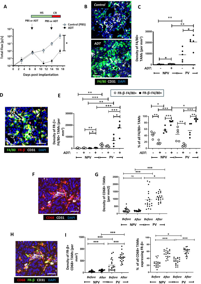
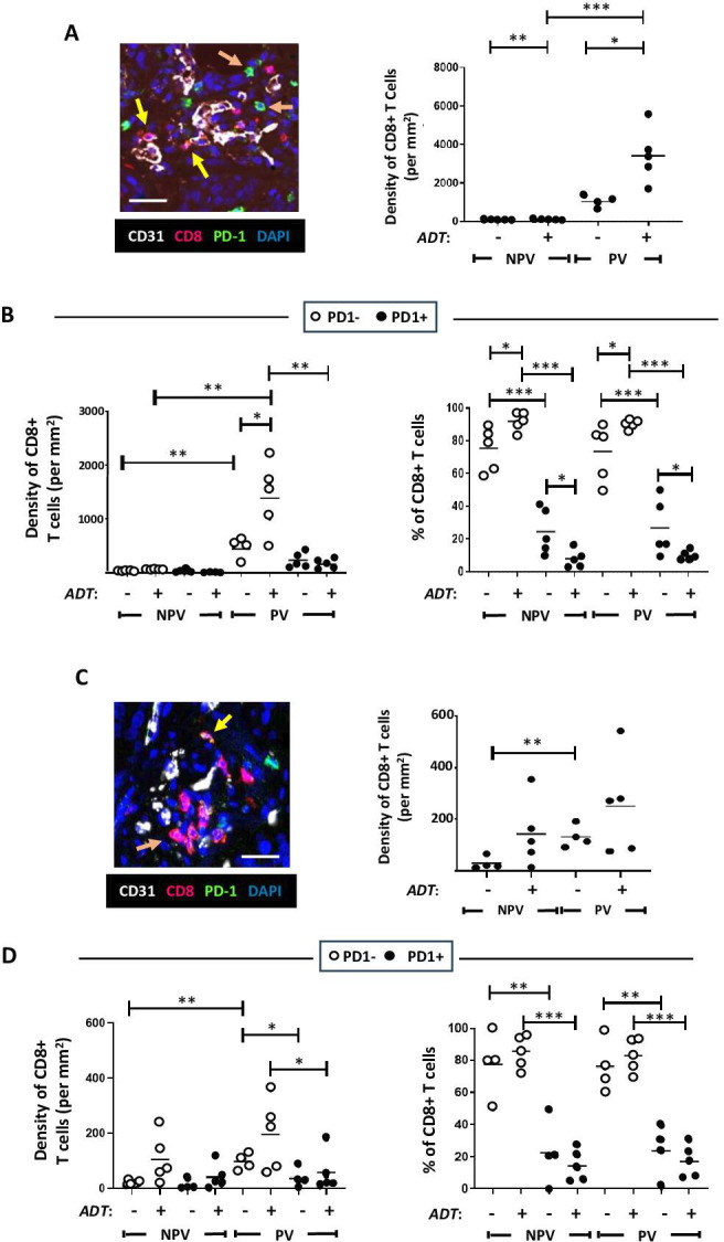
Background: Androgen deprivation therapy (ADT) is a front-line treatment for prostate cancer. In some men, their tumors can become refractory leading to the development of castration-resistant prostate cancer (CRPC). This causes tumors to regrow and metastasize, despite ongoing treatment, and impacts negatively on patient survival. ADT is known to stimulate the accumulation of immunosuppressive cells like protumoral tumor-associated macrophages (TAMs), myeloid-derived suppressor cells and regulatory T cells in prostate tumors, as well as hypofunctional T cells. Protumoral TAMs have been shown to accumulate around tumor blood vessels during chemotherapy and radiotherapy in other forms of cancer, where they drive tumor relapse. Our aim was to see whether such perivascular (PV) TAMs also accumulate in ADT-treated prostate tumors prior to CRPC, and, if so, whether selectively inducing them to express a potent immunostimulant, interferon beta (IFNβ), would stimulate antitumor immunity and delay CRPC.

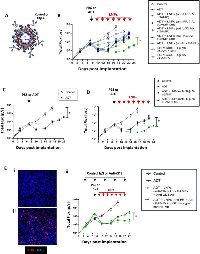
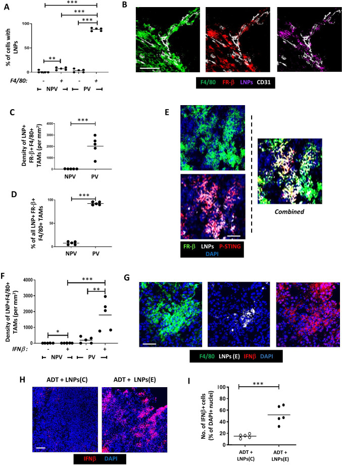
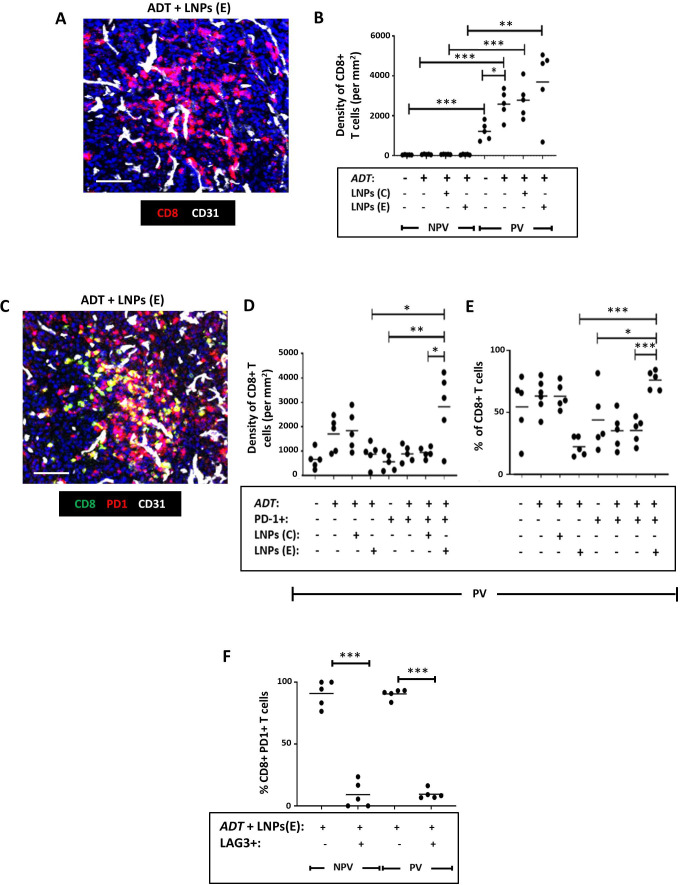
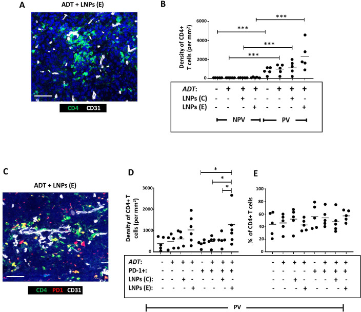
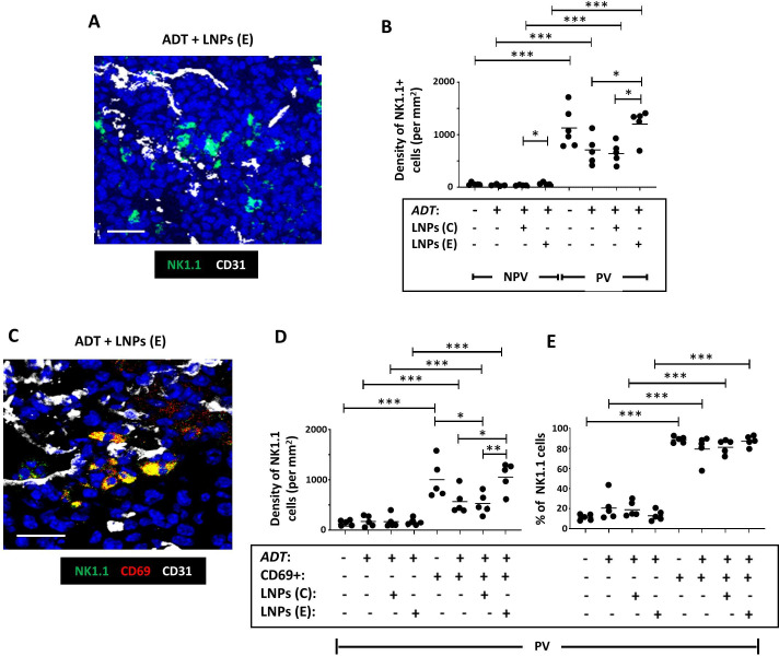
Methods: We used multiplex immunofluorescence to assess the effects of ADT on the distribution and activation status of TAMs, CD8+T cells, CD4+T cells and NK cells in mouse and/or human prostate tumors. We then used antibody-coated, lipid nanoparticles (LNPs) to selectively target a STING agonist, 2'3'-cGAMP (cGAMP), to PV TAMs in mouse prostate tumors during ADT.

Results: TAMs accumulated at high density around blood vessels in response to ADT and expressed markers of a protumoral phenotype including folate receptor-beta (FR-β), MRC1 (CD206), CD169 and VISTA. Additionally, higher numbers of inactive (PD-1-) CD8+T cells and reduced numbers of active (CD69+) NK cells were present in these PV tumor areas. LNPs coated with an antibody to FR-β selectively delivered cGAMP to PV TAMs in ADT-treated tumors, where they activated STING and upregulated the expression of IFNβ. This resulted in a marked increase in the density of active CD8+T cells (along with CD4+T cells and NK cells) in PV tumor areas, and significantly delayed the onset of CRPC. Antibody depletion of CD8+T cells during LNP administration demonstrated the essential role of these cells in delay in CRPC induced by LNPs.

Conclusion: Together, our data indicate that targeting a STING agonist to PV TAMs could be used to extend the treatment window for ADT in prostate cancer.

Keywords: immunotherapy; macrophage; prostate cancer.


Conflict of interest statement

Competing interests: No, there are no competing interests.

Figures

Figure 1
Figure 1. ADT stimulates the PV accumulation of FR-β+ TAMs in mouse (Myc-CaP) and localized, human prostate tumors. (A) Two phases of tumor response to the LHRH antagonist, degarelix, in Myc-CaP tumors: an initial, hormone sensitive (HS) period of tumor growth inhibition followed by the start of castration resistance (CR) when tumors start to regrow. In Myc-CaP tumors (B–E), immunofluorescence staining shows that the density of both PV F4/80+TAMs (B, C)—and the FR-β+ subset of these cells (D, E) increased by the start of CR in ADT-treated tumors. Similar changes occurred in non-PV tumor areas but to a lesser extent than in PV areas. The proportion of F4/80+TAMs expressing FR-β also increased in PV and non-PV areas at this time. Immunofluorescence staining of matched human prostate tumors (F, H) sampled before and after ADT showed that ADT increased the PV density of PV CD68+ tumors (G) and the CD68+TAM subset expressing FR-β (I, left panel). The proportion of CD68+TAMs expressing FR-β rose in PV and non-PV tumor areas after ADT (I, right panel). (NPV=non-PV). Data are presented as means±SEMs. All fluorescence images are of ADT-treated tumors (except the top one in panel B). *p<0.05, **p<0.01, ***p<0.001. Magnification bars=50 µm. ADT, androgen deprivation therapy; LHRH, Luteinising hormone-releasing hormone; PV, perivascular; TAM, tumor-associated macrophage.
Figure 2
Figure 2. ADT stimulates the PV accumulation of PD-1-CD8+T cells in mouse (Myc-CaP) (A, B) and human (C, D) prostate tumors. (A, B). (A) Representative fluorescence images showing the presence of mainly PD-1- CD8+T cells in PV areas of ADT-treated Myc-CaP (A) and human (C) prostate tumors (left panels in both). (A, C) Yellow arrows=PD-1+CD8+T cells, orange arrows=PD-1-CD8+T cells. ADT stimulates the PV accumulation of CD8+T cells (A, C, right panels), which are mainly PD-1- (B, D left panels). The majority of CD8+T cells lack expression of PD-1 across tumors, which increases further after ADT (B, D, right panels). (NPV=non-PV). Data are presented as means±SEMs. Fluorescence images in A, C are from ADT-treated tumors. *p<0.05, **p<0.01, ***p<0.001. Magnification bars=20 µm. ADT, androgen deprivation therapy; PV, perivascular.
Figure 3
Figure 3. LNPs coated with FR-β-antibody target the STING agonist, cGAMP, to PV TAMs and delay CR in ADT-treated Myc-CaP tumors. (A) Design of LNPs used in vivo. The Fc regions of either a FR-β antibody or a control IgG were attached to LNPs containing either an active cGAMP or an inactive version of this (“cGAMP Ctrl”). (B) Tumor growth in mice administered either PBS or ADT alone, or these followed by administration every 2 days of the various forms of LNP listed. (C and D) Various key groups have been selected from (B) and shown separately (for clarity). (C) Tumor growth curves in response to PBS alone (control) versus a single dose of ADT or (D) PBS alone (control), ADT alone, ADT plus FR-β antibody-coated LNPs containing either cGAMP or cGAMP Ctrl. (E) Effect of in vivo administration of (i) an antibody against CD8 or (ii) an isotype-matched control IgG2b on tumor-infiltrating CD8+T cells after ADT plus FR-β antibody-coated LNPs containing cGAMP (magnification bar=50 µm). (iii) Tumor growth curves showing the effect of depleting CD8+T cells on tumor responses to ADT plus by FR-β antibody-coated LNPs containing cGAMP. Data are presented as means±SEMs. *p<0.001 (comparing tumor sizes at sacrifice). ADT, androgen deprivation therapy; LNP, lipid nanoparticle; TAM, tumor-associated macrophage.
Figure 4
Figure 4. Selective delivery of cGAMP to PV FR-β+ TAMs in Myc-CaP tumors results in STING activation and upregulation of IFNβ. Following the administration of ADT plus LNPs: (A). The proportion of cells in PV and non-PV areas bearing LNPs that were F4/80− vs F4/80+. (B) Fluorescently labeled LNPs colocalized with PV FR-β+F4/80+TAMs. (C, D) FR-β+F4/80+TAMs bearing LNPs were only present in PV areas. (E) When LNPs bearing active cGAMP (LNPs(E)) were administered, the expression of active phosphorylated STING (P-STING) could be detected in LNP+FR-β+F4/80+TAMs. This was accompanied by a significant increase in IFNβ detection PV LNP+F4/80+TAMs (ie, in the LNPs(E) group) (E). In this group, IFNβ detection was only detectable in F4/80+TAMs in PV not NPV areas, (F) but often extended beyond LNP+cells, indicating its possible release and uptake by other cells in tumors (G). This did not occur when mice were injected with LNPs bearing inactive cGAMP. (NPV=non-PV). Data are presented as means±SEMs. *p<0.05, **p<0.01, ***p<0.001. Magnification bars=50 µm. ADT, androgen deprivation therapy; LNP, lipid nanoparticle.
Figure 5
Figure 5. Selective delivery of cGAMP to PV FR-β+ TAMs in Myc-CaP tumors reverses the effect of ADT on the activation status of CD8+T cells in Myc-CaP tumors. (A) Representative fluorescence image showing CD8+T cells in a vascularized area of a tumor treated with ADT plus FR-β antibody-coated LNPs containing active cGAMP (“LNPs(E)”). (B) ADT increased the PV accumulation of CD8+T cells (a change that was unaffected by coadministration with FR-β antibody-coated LNPs containing either inactive cGAMP (“LNPs(C)”) or LNPs(E)). (C) Representative fluorescence image showing colocalization of PD-1 and CD8 in a vascularized area of a tumor treated with ADT plus LNPs(E). (D, E) ADT administered with LNPs(E) reversed the PV accumulation of inactive (PD-1-) CD8+T cells induced by ADT alone, and led to an increase in both the PV density (D) and proportion (E) of CD8+T cells expressing PD-1. (F) The majority of PD-1+CD8+ T cells did not express the exhaustion marker, LAG3 following treatment with ADT plus LNPs(E). (NPV=non-PV). Data are presented as means±SEMs. *p<0.05, **p<0.01, ***p<0.001. Magnification bars=50 µm. ADT, androgen deprivation therapy; LNP, lipid nanoparticle.
Figure 6
Figure 6. Selective delivery of cGAMP to PV FR-β+ TAMs in Myc-CaP tumors increased the PV accumulation of PD-1+CD4+ T cells in Myc-CaP tumors. (A) Representative fluorescence image showing CD4+T cells in a vascularized area of a tumor treated with ADT plus FR-β antibody-coated LNPs containing active cGAMP (“LNPs(E)”). (B) CD4+T cells were mainly PV in PBS-treated tumors and this was unaffected by ADT alone or coadministration of ADT with FR-β antibody-coated LNPs containing either inactive cGAMP (“LNPs(C)”) or LNPs(E). (C) Representative fluorescence image showing colocalization of PD-1 and CD4 in a vascularized area of a tumor treated with ADT plus FR-β antibody-coated LNPs containing active cGAMP. (D, E) ADT administered with LNPs(E) resulted in the PV accumulation of PD-1+CD4+ T cells. The proportion of CD4+T cells expressing PD-1 remained unaltered by this treatment. (NPV=non-PV). Data are presented as means±SEMs. *p<0.05, ***p<0.001. Magnification bars=50 µm. ADT, androgen deprivation therapy; LNP, androgen deprivation therapy.
Figure 7
Figure 7. Selective delivery of cGAMP to PV FR-β+ TAMs in Myc-CaP tumors increases the PV density of active NK cells in Myc-CaP tumors. (A) Representative fluorescence image showing NK1.1+ NK cells in a vascularized area of a tumor treated with ADT plus FR-β antibody-coated LNPs containing active cGAMP (“LNPs (E)”). (B) NK cells were mainly PV in PBS-treated tumors and this was unaffected by ADT alone. Coadministration of ADT with LNPs (E) increased both the non-PV and PV density of NK cells compared with ADT alone or ADT plus FR-β antibody-coated LNPs containing inactive cGAMP (“LNPs (C)”). (C) Representative fluorescence image showing colocalization of CD69 and NK1.1 in a vascularized area of a tumor treated with ADT plus LNPs (E). (D, E) ADT administered with LNPs(E) increased the PV density of CD69+ (ie, activated) NK cells compared with ADT alone or ADT+LNPs (C). The majority of PV NK cells expressed CD69 in PBS-treated tumors. This did not change after ADT, with or without LNPs. (NPV=non-PV). Data are presented as means±SEMs. *p<0.05, **p<0.01,***p<0.001. Magnification bars=20 µm. ADT, androgen deprivation therapy; LNP, androgen deprivation therapy.

References

    1. Jamroze A, Chatta G, Tang DG. Androgen receptor (AR) heterogeneity in prostate cancer and therapy resistance. Cancer Lett. 2021;518:1–9. doi: 10.1016/j.canlet.2021.06.006. - DOI - PMC - PubMed
    1. Huang Y, Jiang X, Liang X, et al. Molecular and cellular mechanisms of castration resistant prostate cancer (review) Oncol Lett. 2018;15:6063–76. doi: 10.3892/ol.2018.8123. - DOI - PMC - PubMed
    1. Mansinho A, Macedo D, Fernandes I, et al. Castration-resistant prostate cancer: mechanisms, targets and treatment. Adv Exp Med Biol. 2018;1096:117–33. doi: 10.1007/978-3-319-99286-0_7. - DOI - PubMed
    1. Cattrini C, Zanardi E, Vallome G, et al. Targeting androgen-independent pathways: new chances for patients with prostate cancer? Crit Rev Oncol Hematol. 2017;118:42–53. doi: 10.1016/j.critrevonc.2017.08.009. - DOI - PubMed
    1. Shen Y-C, Ghasemzadeh A, Kochel CM, et al. Combining intratumoral treg depletion with androgen deprivation therapy (ADT): preclinical activity in the myc-cap model. Prostate Cancer Prostatic Dis. 2018;21:113–25. doi: 10.1038/s41391-017-0013-x. - DOI - PMC - PubMed

MeSH terms