. 2024 Jul 25;12(7):e008769.
doi: 10.1136/jitc-2023-008769.

Preclinical activity of allogeneic SLAMF7-specific CAR T-cells (UCARTCS1) in multiple myeloma

Affiliations

Preclinical activity of allogeneic SLAMF7-specific CAR T-cells (UCARTCS1) in multiple myeloma

Charlotte L B M Korst et al. J Immunother Cancer. .

Abstract

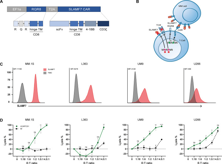
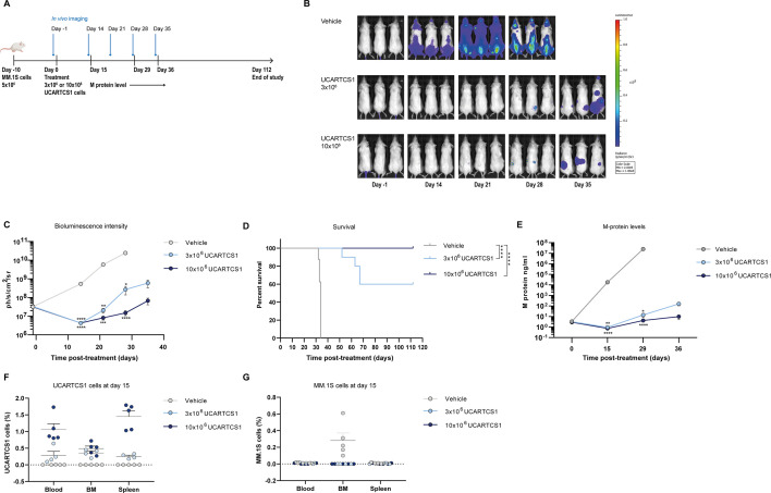
Background: Autologous BCMA-specific CAR T-cell therapies have substantial activity in multiple myeloma (MM). However, due to logistical limitations and BCMAlow relapses, there is a need for alternatives. UCARTCS1 cells are 'off-the-shelf' allogeneic CAR T-cells derived from healthy donors targeting SLAMF7 (CS1), which is highly expressed in MM cells. In this study, we evaluated the preclinical activity of UCARTCS1 in MM cell lines, in bone marrow (BM) samples obtained from MM patients and in an MM mouse model.

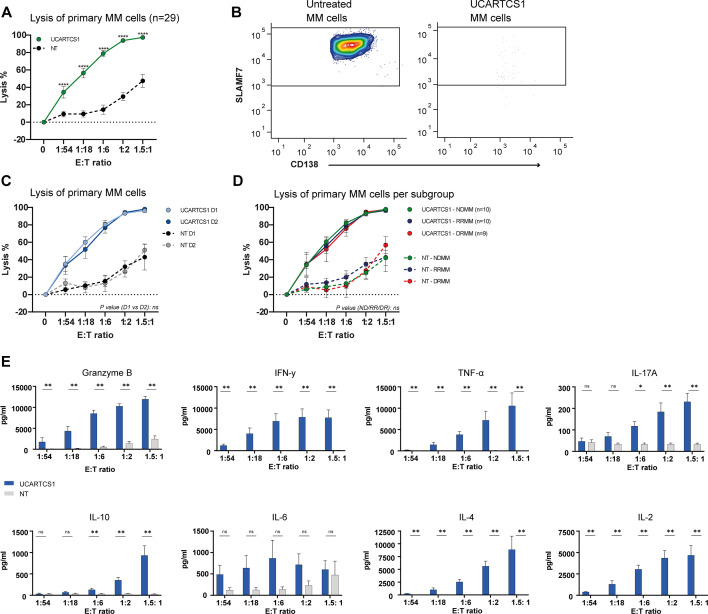
Methods: Luciferase-transduced MM cell lines were incubated with UCARTCS1 cells or control (non-transduced, SLAMF7/TCRαβ double knock-out) T-cells at different effector to target ratios for 24 hours. MM cell lysis was assessed by bioluminescence. Anti-MM activity of UCARTCS1 was also evaluated in 29 BM samples obtained from newly diagnosed patients (n=10), daratumumab-naïve relapsed/refractory patients (n=10) and daratumumab-refractory patients (n=9) in 24-hour flow cytometry-based cytotoxicity assays. Finally, UCARTCS1 activity was assessed in mouse xenograft models.

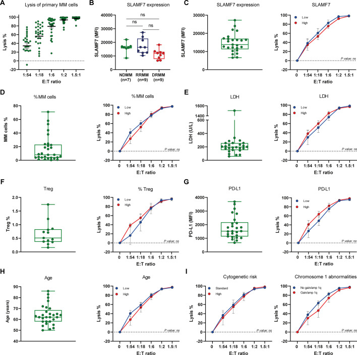
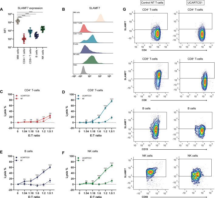
Results: UCARTCS1 cells induced potent CAR-mediated, and dose-dependent lysis of both MM cell lines and primary MM cells. There was no difference in ex vivo activity of UCARTCS1 between heavily pretreated and newly diagnosed patients. In addition, efficacy of UCARTCS1 was not affected by SLAMF7 expression level on MM cells, proportion of tumor cells, or frequency of regulatory T-cells in BM samples obtained from MM patients. UCARTCS1 treatment eliminated SLAMF7+ non-malignant immune cells in a dose-dependent manner, however lysis of normal cells was less pronounced compared to that of MM cells. Additionally, durable anti-MM responses were observed with UCARTCS1 in an MM xenograft model.

Conclusions: These results demonstrate that UCARTCS1 has potent anti-MM activity against MM cell lines and primary MM cells, as well as in an MM xenograft model and support the evaluation of UCARTCS1 in patients with advanced MM.

Keywords: Chimeric antigen receptor - CAR; Immunotherapy; Multiple Myeloma; T cell.


Conflict of interest statement

Competing interests: DLC, IC-S and RG are employees of Cellectis SA. MT has received research support from Kite-Gilead, consultancy compensation from Sangamo Therapeutics and is inventor of licensed patents and patent applications related to the development of CARs and CAR-T cells from iPSC. SZ has received research funding from Celgene, Takeda, and Janssen Pharmaceuticals and serves on advisory boards for Janssen Pharmaceuticals, Sanofi, Celgene, Takeda, Amgen and Oncopeptides. No personal funding. TM has received research support from Janssen Pharmaceuticals, Genmab, Takeda, Novartis and ONK Therapeutics. NWCJvdD has received research support from Janssen Pharmaceuticals, AMGEN, Celgene, Novartis, Cellectis and BMS and serves on advisory boards for Janssen Pharmaceuticals, AMGEN, Celgene, BMS, Takeda, Roche, Novartis, Bayer, Pfizer, Kite Pharma, Merck, Abbvie, Adaptive and Servier, all paid to employer.

Figures

Figure 1
Figure 1. UCARTCS1-mediated lysis of cell lines in vitro. (A) Schematic representation of UCARTCS1 CAR construct containing scFv, CD8-derived hinge and transmembrane domain, 4-1BB, and CD3ζ signaling domains. The CAR is coexpressed with RQR8 as safety feature, by conferring sensitivity to rituximab. (B) UCARTCS1 cell expressing SLAMF7-targeting CAR and RQR8. The TCRαβ receptor is disrupted by knocking out the TRAC gene. Fratricide is prevented by SLAMF7 gene knockout. (C) Flow cytometry histogram overlays depicting SLAMF7 cell surface expression on four MM cell lines (MM.1S, L363, UM9 and U266; red histogram), compared with FMO (fluorescence minus one; gray histogram). SLAMF7 MFI is provided. (D) UCARTCS1-mediated lysis in four MM cell lines. Non-transduced (NT), SLAMF7/TCRαβ double knock-out T-cells were used as control. MM cell lysis was assessed using a 24-hour BLI-based cytotoxicity assay. Data represent mean MM cell lysis±SEM of three independent experiments performed in duplicate. Unpaired t-test was used to calculate significance between both groups. *p<0.05, **p<0.01, ***p<0.001, ****p<0.0001. E:T ratio, effector to target-ratio; MM, multiple myeloma; MFI, median fluorescence intensity; ns, not significant; scFv, single-chain variable fragment; TCR, T-cell receptor
Figure 2
Figure 2. UCARTCS1-mediated lysis of primary MM cells. (A) BM-MNC obtained from 29 patients were incubated with UCARTCS1 cells or control (non-transduced (NT), SLAMF7/TCRαβ double knock-out) T-cells at different E:T ratios for 24 hours, after which the surviving MM cells were enumerated using flow cytometric analysis. Data represent mean±SEM. Mann-Whitney U test was used to calculate significance between both groups. (B) Representative flow cytometry contour plots showing SLAMF7-positive MM cells from a newly diagnosed MM patient, untreated (left) or treated with UCARTCS1 cells (right; E:T ratio of 1.5:1). After exclusion of doublets and dead Fixable Near-IR positive cells, MM cells were identified as CD38+ CD138+ cells. (C) UCARTCS1-mediated lysis of MM cells was similar when using UCARTCS1 cells from two different healthy T-cell donors. UCARTCS1 cells (light blue) and control NT T-cells (black) from donor one were tested with 16 BM samples; UCARTCS1 cells (dark blue) and control NT T-cells (gray) from donor two were tested with 13 BM samples. Mann-Whitney test was used to evaluate significance between both groups. (D) UCARTCS1-mediated lysis of MM cells in patient subgroups according to prior treatment: 10 BM samples from newly diagnosed MM patients (NDMM; green), 10 from daratumumab-naïve RRMM patients (RRMM; blue), and nine from daratumumab-refractory MM patients (DRMM; red). Solid lines represent UCARTCS1 cells and dotted lines control (non-transduced (NT), SLAMF7/TCRαβ double knock-out) T-cells. Data represent mean±SEM. Patient subgroups were compared using the Kruskal-Wallis test. (E) IFN-y, TNF-α, IL-17a, IL-10, IL-6, IL-4, IL-2, and granzyme B were measured in the cell supernatants of BM-MNCs treated with UCARTCS1 (blue) or control (non-transduced (NT), SLAMF7/TCRαβ double knock-out) T-cells (gray) for 24 hours by using a flow cytometry based assay (cytokines) or an ELISA (granzyme B). BM-MNCs were obtained from six DRMM patients. Data represent mean±SEM. Mann-Whitney U test was used to calculate significance between both groups. *p<0.05, **p<0.01, ****p<0.0001. BM-MNCs, bone marrow mononuclear cells; E:T ratio, effector to target-ratio; MM, multiple myeloma; ns, not significant; TCR, T-cell receptor.
Figure 3
Figure 3. Impact of tumor and patient characteristics on UCARTCS1 activity. (A) Shown are the individual UCARTCS1-mediated MM cell lysis values of all 29 MM patients (10 newly diagnosed MM patients (NDMM), 10 daratumumab-naïve RRMM patients (RRMM), and 9 daratumumab-refractory MM patients (DRMM)). In these experiments, BM-MNCs were incubated with UCARTCS1 cells at different E:T ratios for 24 hours, after which the surviving MM cells were enumerated using flow cytometric analysis. Data represent mean±SEM. Each dot represents an individual BM sample. (B) SLAMF7 expression level on MM cells from 7 NDMM, 9 RRMM and 9 DRMM patients, as determined by flow cytometry. Each dot represents an individual sample, with box and whiskers, representing median, 25th–75th percentile, and range. Groups were compared with Kruskal-Wallis test. (C–I) The impact of tumor and patient characteristics on MM cell lysis was assessed by constructing dose-response curves for UCARTCS1-mediated MM cell lysis, according to median SLAMF7 expression level (n=25), median frequency of MM cells (n=29), median LDH level (n=26), median frequency of Tregs (n=13), median PD-L1 expression level (n=26), median age (n=29), cytogenetic risk status (n=27), and presence/absence of chromosome one abnormalities (gain/amp 1q) (n=27). Data represent mean±SEM. (C–I) Blue lines represent samples with a value below or equal to the median value, and red lines samples above the median value. Groups were compared using Mann-Whitney U test. (C–H) Each dot represents an individual sample, with box and whiskers, representing median, 25th–75th percentile, and range. Amp 1q, amplification 1q; BM-MNCs, bone marrow mononuclear cells; E:T ratio, effector to target-ratio; LDH, lactate dehydrogenase; PD-L1, programmed death-ligand 1; RRMM, relapsed/refractory multiple myeloma; TCR, T-cell receptor; Treg, regulatory T-cell; ns, not significant.
Figure 4
Figure 4. Effect of UCARTCS1 on non-malignant immune cells. (A) SLAMF7 expression on MM cells was compared with SLAMF7 expression on CD4+ T-cells, CD8+ T-cells, B cells and NK cells in 25 BM samples from MM patients (the same patients’ BM samples were also used in killing assays) by Wilcoxon matched-pairs test. Each dot represents an individual sample, with box and whiskers, representing median, 25th–75th percentile, and range. (B) Representative flow cytometry histogram overlays depicting SLAMF7 expression on MM cells, CD4+ T-cells, CD8+ T-cells, B cells and NK cells in a BM sample from a newly diagnosed MM patient. Fluorescence minus one (FMO) is depicted in gray (gated on MM cells). (C–F) BM-MNCs obtained from 29 MM patients were incubated with UCARTCS1 cells or control (non-transduced (NT), SLAMF7/TCRαβ double knock-out) T-cells at different E:T ratios for 24 hours after which surviving CD4+ T-cells, CD8+ T-cells, B cells and NK cells were enumerated using flow cytometric analysis. Solid lines represent UCARTCS1 cells and dotted lines control NT T-cells. Data represent mean±SEM. Groups were compared using Mann-Whitney U test. (G) Representative flow cytometry contour plots showing CD4+ T-cells, CD8+ T-cells, B cells and NK cells from a newly diagnosed MM patient after treatment with control (non-transduced (NT), SLAMF7/TCRαβ double knock-out) T-cells (left) or UCARTCS1 cells (right) (E:T ratio of 1.5 : 1). *p<0.05, **p<0.01, ***p<0.001, ****p<0.0001. BM-MNCs, bone marrow mononuclear cells; E:T ratio, effector to target-ratio; MM, multiple myeloma; ns, not significant; TCR, T-cell receptor.
Figure 5
Figure 5. In vivo activity of UCARTCS1. (A) NSG mice injected with LUC-GFP-transduced MM.1S (iv) on day −10 and treated with a single dose of 3×10⁶ UCARTCS1 cells, or 10×106 UCARTCS1 cells, or vehicle control (on day 0). (B, C) BLI readout performed on day −1, day 14, day 21, day 28, and day 35 to evaluate tumor progression in the different treatment groups. Both treatment groups were compared with vehicle control using Kruskal Wallis test. (D) Survival curves of mice treated with a single dose of 3×10⁶ UCARTCS1 cells, a single dose of 10×106 UCARTCS1 cells, or vehicle control. Groups were compared using logrank test. (E) M-protein levels (ng/mL) assessed in mice at day 15, day 29 and day 36. Both treatment groups were compared with vehicle control using Kruskal-Wallis test. Data represent mean±SEM. (F, G) Shown are percentages of UCARTCS1 cells and MM.1S cells in the blood, BM and spleen of the subset of mice sacrificed at day 15 (4–5 mice per group). Presence of UCARTCS1 and MM.1S cells was assessed using flow cytometric analysis. UCARTCS1 cells were identified by the expression of human CD45 (hCD45) and by negativity for GFP. MM.1S-LUC-GFP tumor cells were identified as GFP+/hCD45- cells. Data represent mean±SEM. Each dot represents an individual mouse. *p<0.05, **p<0.01, ***p<0.001, ****p<0.0001. BLI, bioluminescence imaging; LUC, luciferase; MM, multiple myeloma; ns, not significant; NSG, NOD SCID gamma mice.

Similar articles

Cited by

References

    1. Mateos M-V, Weisel K, De Stefano V, et al. LocoMMotion: a prospective, non-interventional, multinational study of real-life current standards of care in patients with relapsed/refractory multiple myeloma (RRMM) receiving ≥3 prior lines of therapy. J Clin Oncol. 2021;39:8041. doi: 10.1200/JCO.2021.39.15_suppl.8041. - DOI - PMC - PubMed
    1. Gandhi UH, Cornell RF, Lakshman A, et al. Outcomes of patients with multiple myeloma refractory to CD38-targeted monoclonal antibody therapy. Leukemia. 2019;33:2266–75. doi: 10.1038/s41375-019-0435-7. - DOI - PMC - PubMed
    1. Munshi NC, Anderson LD, Jr, Shah N, et al. Idecabtagene vicleucel in relapsed and refractory multiple myeloma. N Engl J Med. 2021;384:705–16. doi: 10.1056/NEJMoa2024850. - DOI - PubMed
    1. Berdeja JG, Madduri D, Usmani SZ, et al. Ciltacabtagene autoleucel, a B-cell maturation antigen-directed chimeric antigen receptor T-cell therapy in patients with relapsed or refractory multiple myeloma (CARTITUDE-1): a phase 1b/2 open-label study. Lancet. 2021;398:314–24. doi: 10.1016/S0140-6736(21)00933-8. - DOI - PubMed
    1. Martin T, Usmani SZ, Berdeja JG, et al. Ciltacabtagene autoleucel, an anti-B-cell maturation antigen chimeric antigen receptor T-cell therapy, for relapsed/refractory multiple myeloma: CARTITUDE-1 2-year follow-up. J Clin Oncol. 2023;41:1265–74. doi: 10.1200/JCO.22.00842. - DOI - PMC - PubMed

Substances