. 2024 Jul 27;24(1):279.
doi: 10.1186/s12866-024-03418-x.

The transcriptional regulator Fur modulates the expression of uge, a gene essential for the core lipopolysaccharide biosynthesis in Klebsiella pneumoniae

Affiliations

The transcriptional regulator Fur modulates the expression of uge, a gene essential for the core lipopolysaccharide biosynthesis in Klebsiella pneumoniae

José Júlio Muner et al. BMC Microbiol. .

Abstract

Background: Klebsiella pneumoniae is a Gram-negative pathogen that has become a threat to public health worldwide due to the emergence of hypervirulent and multidrug-resistant strains. Cell-surface components, such as polysaccharide capsules, fimbriae, and lipopolysaccharides (LPS), are among the major virulence factors for K. pneumoniae. One of the genes involved in LPS biosynthesis is the uge gene, which encodes the uridine diphosphate galacturonate 4-epimerase enzyme. Although essential for the LPS formation in K. pneumoniae, little is known about the mechanisms that regulate the expression of uge. Ferric uptake regulator (Fur) is an iron-responsive transcription factor that modulates the expression of capsular and fimbrial genes, but its role in LPS expression has not yet been identified. This work aimed to investigate the role of the Fur regulator in the expression of the K. pneumoniae uge gene and to determine whether the production of LPS by K. pneumoniae is modulated by the iron levels available to the bacterium.

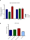
Results: Using bioinformatic analyses, a Fur-binding site was identified on the promoter region of the uge gene; this binding site was validated experimentally through Fur Titration Assay (FURTA) and DNA Electrophoretic Mobility Shift Assay (EMSA) techniques. RT-qPCR analyses were used to evaluate the expression of uge according to the iron levels available to the bacterium. The iron-rich condition led to a down-regulation of uge, while the iron-restricted condition resulted in up-regulation. In addition, LPS was extracted and quantified on K. pneumoniae cells subjected to iron-replete and iron-limited conditions. The iron-limited condition increased the amount of LPS produced by K. pneumoniae. Finally, the expression levels of uge and the amount of the LPS were evaluated on a K. pneumoniae strain mutant for the fur gene. Compared to the wild-type, the strain with the fur gene knocked out presented a lower LPS amount and an unchanged expression of uge, regardless of the iron levels.

Conclusions: Here, we show that iron deprivation led the K. pneumoniae cells to produce higher amount of LPS and that the Fur regulator modulates the expression of uge, a gene essential for LPS biosynthesis. Thus, our results indicate that iron availability modulates the LPS biosynthesis in K. pneumoniae through a Fur-dependent mechanism.

Keywords: Klebsiella pneumoniae; Gene expression analyses; Iron-regulatory proteins; Lipopolysaccharides; Transcriptional regulation.


Conflict of interest statement

The authors declare no competing interests.

Figures

Fig. 1
Fig. 1
The promoter region of uge harbors a functional Fur binding box validated by FURTA and EMSA. A Partial nucleotide sequence of the 5’-upstream region of uge showing the initial codon (ATG, underlined) and the ribosome binding site (RBS, highlighted in grey). Uppercase nucleotides on RBS are identical to the K. pneumoniae RBS proposed by Kim et al. [24]. The transcription initiation site of uge is indicated at position + 1 (underlined Adenine in red). BPROM program predicted the -35 (in green) and -10 (in yellow) domains of the Sigma factor RpoD. The putative Fur binding box, identified 154 nucleotides upstream of the uge start codon, is highlighted in blue. The functionality of this Fur box was experimentally validated by FURTA and EMSA. B FURTA, E. coli H1717 transformed with a functional Fur box will appear red on MacConkey agar plates (positive control, Lac+ phenotype), whereas H1717 transformed with a non-functional Fur box will appear colorless (negative control, Lac phenotype). FURTA validated the Fur box from uge, as indicated by the red colonies of E. coli H1717 transformed with the Fur box sequence from uge (FURTA-positive phenotype). C EMSA, a mobility shift was observed when the DNA probes containing the Fur box from uge were incubated with K. pneumoniae purified His-Fur protein in the presence of divalent cation (lane 2), compared to the DNA probes alone (without the addition of His-Fur protein, lane 1). The mobility shift of the probes was abolished under divalent cation-free conditions (in the presence of the chelator EDTA, lane 3). No mobility shift was observed with the DNA probe without the Fur box sequence from uge (the negative control). Open arrowhead indicates the DNA probes alone, while closed arrowhead indicates the mobility shift corresponding to the Fur/DNA complexes. Full-length gels are presented in Supplementary Figure S1
Fig. 2
Fig. 2
Iron levels modulate the expression of uge and the amounts of LPS produced by K. pneumoniae. A RT-qPCR analyses showed down-regulation and up-regulation of uge expression, respectively, under iron-replete (FeSO4) and iron-limiting (dipyridyl) conditions in the ATCC 10031 wild-type (wt) strain. The strain mutant for the fur gene has unchanged expressions of uge in all the conditions tested. B Iron-limited conditions led the K. pneumoniae cells to produce higher amounts of LPS when compared to the control and iron-replete (FeSO4) conditions. The iron-rich condition presented no change in LPS production compared to the control condition. The fur mutant strain produces less LPS than the ATCC 10031 strain, regardless of the culture conditions in which the strain is submitted. Different letters above the graphical bars indicate statistically significant differences ( p-value < 0.05)

Similar articles

Cited by

References

    1. Podschun R, Ullmann U. Klebsiella spp. as nosocomial pathogens: epidemiology, taxonomy, typing methods, and pathogenicity factors. Clin Microbiol Rev. 1998;11:589–603. 10.1128/CMR.11.4.589 - DOI - PMC - PubMed
    1. Paczosa MK, Mecsas J. Klebsiella pneumoniae: going on the offense with a strong defense. Microbiol Mol Biol Rev. 2016;80:629–61. 10.1128/MMBR.00078-15 - DOI - PMC - PubMed
    1. Lee C-R, Lee JH, Park KS, Jeon JH, Kim YB, Cha C-J, et al. Antimicrobial resistance of hypervirulent Klebsiella pneumoniae: epidemiology, hypervirulence-associated determinants, and resistance mechanisms. Front Cell Infect Microbiol. 2017;7:7. 10.3389/fcimb.2017.00483 - DOI - PMC - PubMed
    1. Rodriguez-Villar S, Fife A, Baldwin C, Warne RR. Antibiotic-resistant hypervirulent Klebsiella pneumoniae causing community- acquired liver abscess: an emerging disease. Oxf Med Case Reports. 2019;2019:omz032. 10.1093/omcr/omz032 - DOI - PMC - PubMed
    1. Choby JE, Howard-Anderson J, Weiss DS. Hypervirulent Klebsiella pneumoniae – clinical and molecular perspectives. J Intern Med. 2020;287:283–300. 10.1111/joim.13007 - DOI - PMC - PubMed

MeSH terms

LinkOut - more resources