. 2024 Jul 17;16(14):2564.
doi: 10.3390/cancers16142564.

Non-Invasive 3D Breast Tumor Localization: A Viable Alternative to Invasive Tumor Marking

Affiliations

Non-Invasive 3D Breast Tumor Localization: A Viable Alternative to Invasive Tumor Marking

Dragana Bjelica et al. Cancers (Basel). .

Abstract

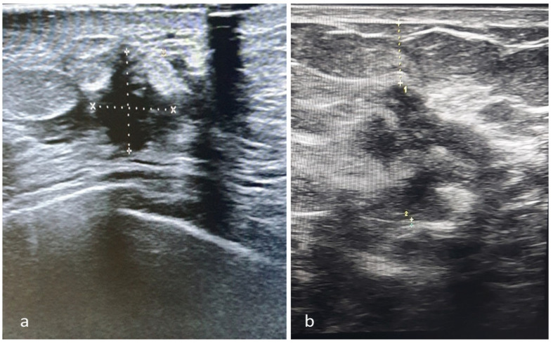
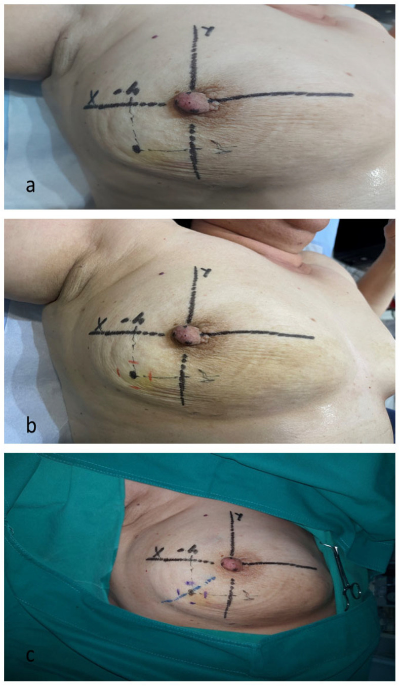
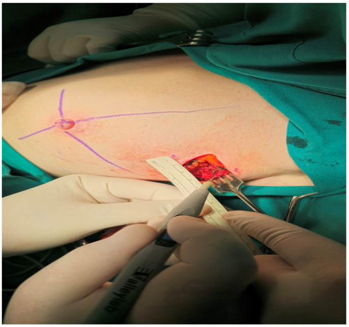
Background: We present a detailed description and the preliminary results of our original technique for non-invasive three-dimensional tumor localization in the breast, which was created as an alternative to standard invasive tumor marking before neoadjuvant systemic therapy (NAST), aiming to enable adequate surgery after complete tumor regression. Methods: A detailed description of the technique is provided in the main text. The technique's feasibility and precision were assessed in a single-arm, prospective study based on the histological parameters of the adequacy and rationality of the excision of completely regressed tumor beds. Results: Out of 94 recruited patients, 15 (16%) were deemed unsuitable, mainly due to the tumors' inadequate ultrasound visibility. Among the 79 processed patients, 31 (39%) had complete clinical regression after NAST and were operated on using our technique. The histological parameters of surgical precision (signs of tumor regression: 24/31; microscopic cancer residues: 7/31) were verified in all excised specimens (100% precision). There were no positive margins in seven cases with microscopic residues, indicating our technique's capacity to enable oncologically safe post-NAST surgery. Conclusions: The proposed technique is feasible and satisfactorily accurate in determining the location of regressed tumors, thus representing an alternative to invasive tumor marking, especially in surgical centers lacking trained staff and equipment for invasive marking. The technique's limitations are mainly related to the inadequate ultrasound visibility of the tumor.

Keywords: early breast cancer; marking techniques; neoadjuvant systemic therapy; pathological complete response; surgical excision; tumor positioning; ultrasound.


Conflict of interest statement

The authors declare no conflicts of interest.

Figures

Figure 1
Figure 1
Drawing the horizontal and vertical axes on the skin of the breast. (a) The horizontal axis is drawn in a line connecting the nipples of both breasts. (b) The vertical axis is drawn at right angles to the horizontal axis. (c) A coordinate system of horizontal and vertical axes drawn on the skin of the breast, intersecting at the nipple.
Figure 2
Figure 2
Determining of the projection of the central point of the tumor on the skin. (a) Ultrasound probe placed orthogonally to the surface of the skin above the tumor; the largest section of the tumor is in the center of the ultrasound screen. (b,c) The distances from the center of the ultrasound probe to the corresponding axes drawn on the skin, measured with a ruler; the long axis of the probe in both positions must be absolutely parallel with the “x” and “y” axes.
Figure 3
Figure 3
The ultrasound measurements. (a) Largest horizontal (craniocaudal and mediolateral) and vertical diameters of the tumor. (b) Distances of the superficial margin of the tumor–skin (1) and the deepest margin–pectoral fascia (2).
Figure 4
Figure 4
Planning of the surgical excision of the tumor bed based on the tumor positioning records. (a) Central point of the tumor drawn based on known coordinates. (b) Red dashes indicate tumor margins in the cranio-caudal and medio-lateral directions. (c) The blue dotted line indicates the skin incision line.
Figure 5
Figure 5
Measurement of spatial relationships by sterile ruler during the operation.

References

    1. Scholl S.M., Asselain B., Palangie T., Dorval T., Jouve M., Giralt E.G., Vilcoq J., Durand J.C., Pouillart P. Neoadjuvant chemotherapy in operable breast cancer. Eur. J. Cancer Clin. Oncol. 1991;27:1668–1671. doi: 10.1016/0277-5379(91)90442-G. - DOI - PubMed
    1. Fisher B., Bryant J., Wolmark N., Mamounas E., Brown A., Fisher E.R., Wickerham D.L., Begovic M., DeCillis A., Robidoux A., et al. Effect of preoperative chemotherapy on the outcome of women with operable breast cancer. J. Clin. Oncol. 1998;16:2672–2685. doi: 10.1200/JCO.1998.16.8.2672. - DOI - PubMed
    1. von Minckwitz G., Huang C.-S., Mano M.S., Loibl S., Mamounas E.P., Untch M., Wolmark N., Rastogi P., Schneeweiss A., Redondo A., et al. Trastuzumab emtansine for residual invasive HER2-positive breast cancer. N. Engl. J. Med. 2019;380:617–628. doi: 10.1056/NEJMoa1814017. - DOI - PubMed
    1. Masuda N., Lee S.-J., Ohtani S., Im Y.-H., Lee E.-S., Yokota I., Kuroi K., Im S.A., Park B.W., Kim S.B., et al. Adjuvant capecitabine for breast cancer after preoperative chemotherapy. N. Engl. J. Med. 2017;376:2147–2159. doi: 10.1056/NEJMoa1612645. - DOI - PubMed
    1. Pfob A., Dubsky P. The underused potential of breast conserving therapy after neoadjuvant system treatment—Causes and solutions. Breast. 2023;67:110–115. doi: 10.1016/j.breast.2023.01.008. - DOI - PMC - PubMed

LinkOut - more resources