. 2024 Jul 7;13(7):631.
doi: 10.3390/antibiotics13070631.

New Fe3O4-Based Coatings with Enhanced Anti-Biofilm Activity for Medical Devices

Affiliations

New Fe3O4-Based Coatings with Enhanced Anti-Biofilm Activity for Medical Devices

Ioana Adelina Pirușcă et al. Antibiotics (Basel). .

Abstract

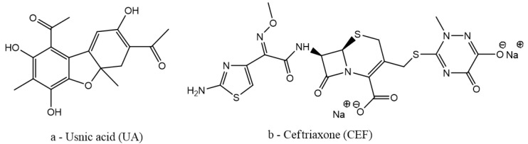
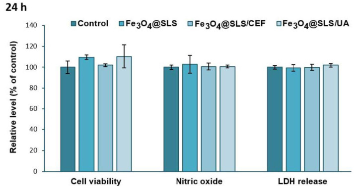
With the increasing use of invasive, interventional, indwelling, and implanted medical devices, healthcare-associated infections caused by pathogenic biofilms have become a major cause of morbidity and mortality. Herein, we present the fabrication, characterization, and in vitro evaluation of biocompatibility and anti-biofilm properties of new coatings based on Fe3O4 nanoparticles (NPs) loaded with usnic acid (UA) and ceftriaxone (CEF). Sodium lauryl sulfate (SLS) was employed as a stabilizer and modulator of the polarity, dispersibility, shape, and anti-biofilm properties of the magnetite nanoparticles. The resulting Fe3O4 functionalized NPs, namely Fe3O4@SLS, Fe3O4@SLS/UA, and Fe3O4@SLS/CEF, respectively, were prepared by co-precipitation method and fully characterized by XRD, TEM, SAED, SEM, FTIR, and TGA. They were further used to produce nanostructured coatings by matrix-assisted pulsed laser evaporation (MAPLE) technique. The biocompatibility of the coatings was assessed by measuring the cell viability, lactate dehydrogenase release, and nitric oxide level in the culture medium and by evaluating the actin cytoskeleton morphology of murine pre-osteoblasts. All prepared nanostructured coatings exhibited good biocompatibility. Biofilm growth inhibition ability was tested at 24 h and 48 h against Staphylococcus aureus and Pseudomonas aeruginosa as representative models for Gram-positive and Gram-negative bacteria. The coatings demonstrated good biocompatibility, promoting osteoblast adhesion, migration, and growth without significant impact on cell viability or morphology, highlighting their potential for developing safe and effective antibacterial surfaces.

Keywords: biofilm inhibition; ceftriaxone; coatings; hydrophobic nanoparticles; nosocomial infections; sodium lauryl sulfate; usnic acid.


Conflict of interest statement

The authors declare no conflicts of interest.

Figures

Figure 1
Figure 1
Chemical structures of the antimicrobial agents.
Figure 2
Figure 2
XRD diffractogram of Fe3O4@SLS.
Figure 3
Figure 3
TEM (a,b) micrographs, SAED pattern (c), and size distribution (d) of Fe3O4@SLS NPs.
Figure 4
Figure 4
Combined TGA-DSC thermogram of pristine Fe3O4 NPs.
Figure 5
Figure 5
Combined TGA-DSC thermogram of core/shell Fe3O4@SLS NPs.
Figure 6
Figure 6
FT-IR spectra of (a1a4) Fe3O4@SLS, (b1b4) Fe3O4@SLS/UA, and (c1c4) Fe3O4@SLS/CEF at (a2,b2,c2) F = 200 mJ/cm2, (a3,b3,c3) F = 300 mJ/cm2, and (a4,b4,c4) F = 400 mJ/cm2 laser fluences and (a1,b1,c1) dropcast.
Figure 7
Figure 7
IR maps of Fe3O4@SLS created based on –CH2– and S–O groups at surface intensity distributions of (a1a4) 2916 cm−1 and (b1b4) 1107 cm−1 for (a1,b1) dropcast and coatings deposited at (a2,b2) 200 mJ/cm2, (a3,b3) 300 mJ/cm2, and (a4,b4) 400 mJ/cm2 laser fluences.
Figure 8
Figure 8
IR maps of Fe3O4@SLS/UA created based on C=O and S–O groups at surface intensity distributions of (a1a4) 1697 cm−1 and (b1b4) is 1107 cm−1 for (a1,b1) dropcast and coatings deposited at (a2,b2) 200 mJ/cm2, (a3,b3) 300 mJ/cm2, and (a4,b4) 400 mJ/cm2 laser fluences.
Figure 9
Figure 9
IR maps of Fe3O4@SLS/CEF created based on C=O and S–O groups at surface intensity distributions of (a1a4) 1658 cm−1 and (b1b4) 1107 cm−1 for (a1,b1) dropcast and coatings deposited at (a2,b2) 200 mJ/cm2, (a3,b3) 300 mJ/cm2, and (a4,b4) 400 mJ/cm2 laser fluences.
Figure 10
Figure 10
SEM micrographs of Fe3O4@SLS (a1,b1,c1,d1), Fe3O4@SLS/UA (a2,b2,c2,d2), and Fe3O4@SLS/CEF (a3,b3,c3,d3) plan-view (a1a3,b1b3,c1c3) and cross-sections (d1d3) recorded at various magnifications.
Figure 11
Figure 11
Biocompatibility of Fe3O4@SLS, Fe3O4@SLS/CEF, and Fe3O4@SLS/UA coatings, as shown by cell viability (MTT assay), NO level, and LDH release after 24 h of exposure on MC3T3-E1 murine cells. All results were calculated as mean ± standard deviations of three different replicates and expressed relative to control.
Figure 12
Figure 12
Biocompatibility of Fe3O4@SLS, Fe3O4@SLS/CEF, and Fe3O4@SLS/UA coatings, as shown by cell viability (MTT assay), NO level, and LDH release after 72 h of exposure on MC3T3-E1 murine cells. All results were calculated as mean ± standard deviations of three different replicates and expressed relative to control.
Figure 13
Figure 13
Actin cytoskeleton organization of MC3T3-E1 murine cells after 24 and 72 h of incubation with Fe3O4@SLS, Fe3O4@SLS/CEF, and Fe3O4@SLS/UA coatings. F-actin (green) was labeled with phalloidin-fluorescein isothiocyanate (FITC), and nuclei (blue) were counterstained with 4′,6-diamidino-2-phenylindole dihydrochloride (DAPI). Magnification (20× objective).
Figure 14
Figure 14
Evaluation of S. aureus biofilm growth after 24/48 h of incubation with Fe3O4@SLS, Fe3O4@SLS/CEF, and Fe3O4@SLS/UA coatings vs. control (one-way ANOVA, when comparing samples vs. control * p < 0.05; ** p < 0.001).
Figure 15
Figure 15
Evaluation of P. aeruginosa biofilm growth after 24/48 h of incubation with Fe3O4@SLS, Fe3O4@SLS/CEF, and Fe3O4@SLS/UA coatings vs. control (one-way ANOVA, when comparing samples vs. control * p < 0.05; ** p < 0.001).

References

    1. Suetens A., Latour K., Kärki T., Ricchizzi E., Kinross P., Moro M.L., Jans B., Hopkins S., Hansen S., Lyytikäinen O., et al. Prevalence of healthcare-associated infections, estimated incidence and composite antimicrobial resistance index in acute care hospitals and long-term care facilities: Results from two European point prevalence surveys, 2016 to 2017. Eurosurveillance. 2018;23:1800516. doi: 10.2807/1560-7917.ES.2018.23.46.1800516. - DOI - PMC - PubMed
    1. Balaure P.C., Grumezescu A.M. Recent Advances in Surface Nanoengineering for Biofilm Prevention and Control. Part I: Molecular Basis of Biofilm Recalcitrance. Passive Anti-Biofouling Nanocoatings. Nanomaterials. 2020;10:1230. doi: 10.3390/nano10061230. - DOI - PMC - PubMed
    1. Balaure P.C., Grumezescu A.M. Recent Advances in Surface Nanoengineering for Biofilm Prevention and Control. Part II: Active, Combined Active and Passive, and Smart Bacteria-Responsive Antibiofilm Nanocoatings. Nanomaterials. 2020;10:1527. doi: 10.3390/nano10081527. - DOI - PMC - PubMed
    1. Cai S., Wu C., Yang W., Liang W., Yu H., Liu L. Recent advance in surface modification for regulating cell adhesion and behaviors. Nanotechnol. Rev. 2020;9:971–989. doi: 10.1515/ntrev-2020-0076. - DOI
    1. Wei J., Yoshinari M., Takemoto S., Hattori M., Kawada E., Liu B., Oda Y. Adhesion of mouse fibroblasts on hexamethyldisiloxane surfaces with wide range of wettability. J. Biomed. Mater. Res. Part B. Appl. Biomater. 2007;81:66–75. doi: 10.1002/jbm.b.30638. - DOI - PubMed

LinkOut - more resources