. 2024 Jul 19;14(14):2110.
doi: 10.3390/ani14142110.

Comparison of MMP-2, MMP-9, COX-2, and PGP Expression in Feline Injection-Site and Feline Noninjection-Site Sarcomas-Pilot Study

Affiliations

Comparison of MMP-2, MMP-9, COX-2, and PGP Expression in Feline Injection-Site and Feline Noninjection-Site Sarcomas-Pilot Study

Agata Wojtkowska et al. Animals (Basel). .

Abstract

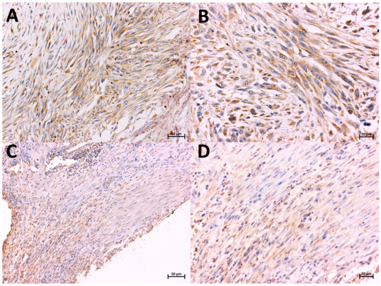
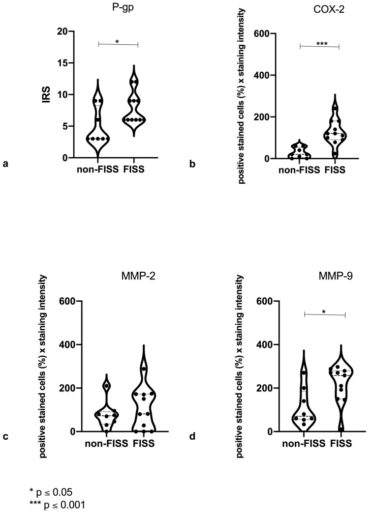
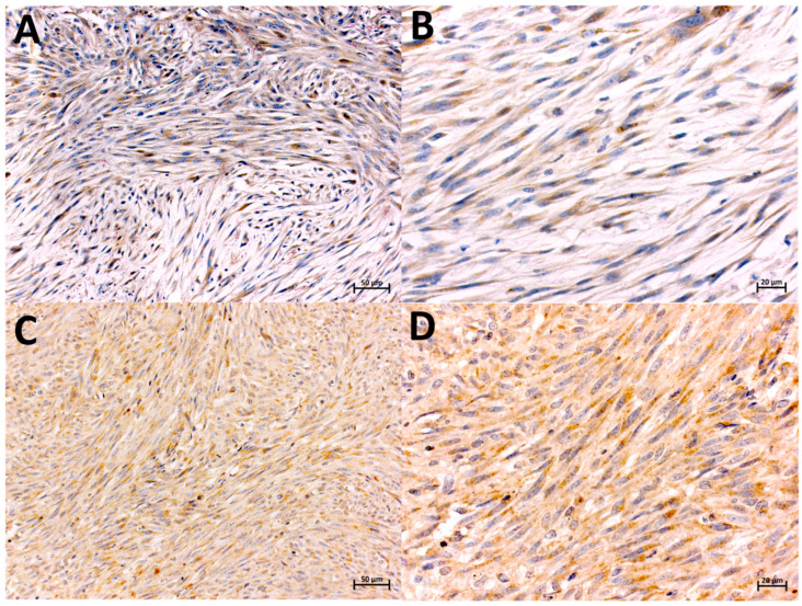
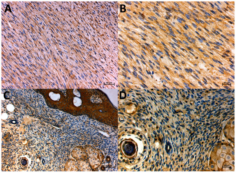
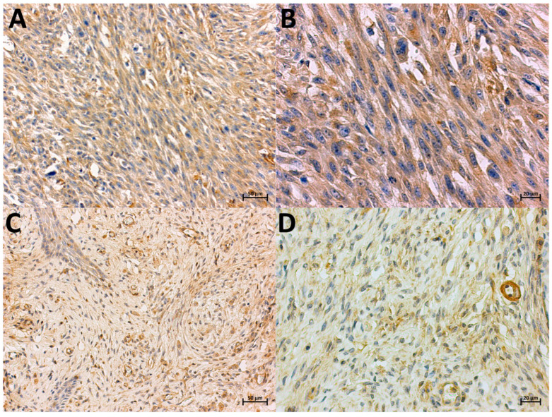
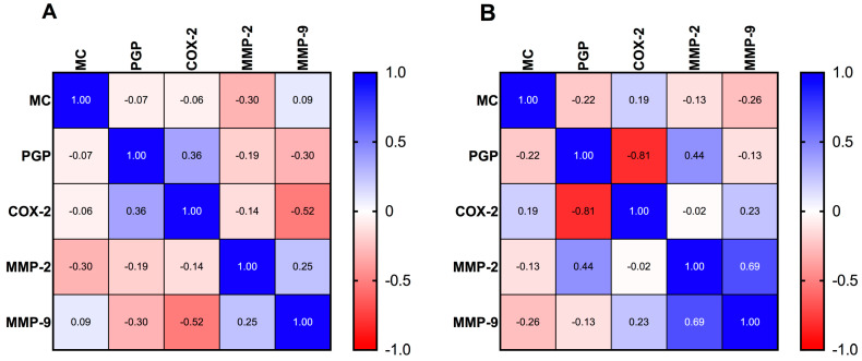
Feline injection-site sarcomas (FISSs) are aggressive neoplasms that have been associated mostly with vaccination. Feline noninjection-site sarcomas (non-FISSs) are less frequently observed in cats and may arise in any anatomic site. This study aimed to determine the differences in the expression of the selected proteins (matrix metalloproteinase-2 (MMP-2), matrix metalloproteinase-9 (MMP-9), cyclooxygenase-2 (COX-2), and P-glycoprotein (PGP)) and their correlation with the mitotic count in FISS and non-FISS, in order to characterize their immunohistochemical features. A preliminary study of eleven samples of FISS and eight samples of non-FISS was performed using immunohistochemistry. Among all the tested sarcomas, 80.4% of the tumors were positive for COX-2, 90.2% were positive for MMP-9, and 100% were positive for PGP. The results showed that the expressions of COX-2, MMP-9, and PGP were significantly higher in FISS than in non-FISS (COX-2-p ≤ 0.001; MMP-9-p ≤ 0.05; and PGP-p ≤ 0.05). A Spearman rank correlation analysis showed a moderate negative correlation between the expression of COX-2 and MMP-9 in FISS (r = -0.52). A strong negative correlation between COX-2 and PGP (r = -0.81), a moderate positive correlation between MMP-2 and MMP-9 (r = +0.69), and a moderate negative correlation between MMP-2 and PGP (r = -0.44) were observed in non-FISS. In summary, our study presents the immunohistochemical profile of the proteins involved with inflammation and carcinogenesis in FISS and non-FISS, which can contribute to expanding the knowledge of tumor biology.

Keywords: P-glycoprotein; cyclooxygenase-2; feline injection-site sarcoma; fibrosarcoma; immunohistochemistry; matrix metalloprotease.


Conflict of interest statement

The authors declare no potential conflicts of interest with respect to the research, authorship, and/or publication of this article.

Figures

Figure 1
Figure 1
Localization of COX-2-expressing cells in FISS (A,B) and non-FISS (C,D), immunohistochemically stained. Brown color indicates positive staining. Positive immunostaining for COX-2 in FISS, intensity 3, 90% of positive cells, magnification 20× and 40×, respectively (A,B). Positive immunostaining for COX-2 in non-FISS, intensity 1, 60% of positive cells, magnification 20× and 40×, respectively (C,D).
Figure 2
Figure 2
Statistical analysis of P-glycoprotein (PGP) (a), cyclooxygenase 2 (COX-2) (b), matrix metalloproteinase-2 (MMP-2) (c), and matrix metalloproteinase-9 (MMP-9) (d) expressions in evaluated groups. (a) Significant (*) (p ≤ 0.05) difference in PGP expression with feline injection-site sarcoma (FISS) and with feline noninjection-site sarcoma (non-FISS). (b) Significantly higher (***) (p ≤ 0.001) expression of COX-2 with FISS and non-FISS. (c) No significant difference in MMP-2 expression with FISS and non-FISS. (d) Significantly (*) (p ≤ 0.05) higher expression of MMP-9 with FISS than with non-FISS.
Figure 3
Figure 3
Localization of MMP-2-expressing cells in FISS (A,B) and non-FISS (C,D), immunohistochemically stained. Brown color indicates positive staining. Positive immunostaining for MMP-2 in FISS, intensity 2, 87% of positive cells, magnification 20× and 40×, respectively (A,B). Positive immunostaining for MMP-2 in non-FISS, intensity 1, 90% of positive cells, magnification 20× and 40×, respectively (C,D).
Figure 4
Figure 4
Localization of the MMP-9-expressing cells in FISS (A,B) and non-FISS (C,D), immunohistochemically stained. Brown color indicates positive staining. Positive immunostaining for MMP-9 in FISS, intensity 3, 96% of positive cells, magnification 20× and 40×, respectively (A,B). Positive immunostaining for MMP-9 in non-FISS, intensity 3, 90% of positive cells, magnification 20× and 40×, respectively (C,D).
Figure 5
Figure 5
Localization of PGP-expressing cells in FISS (A,B) and non-FISS (C,D), immunohistochemically stained. Brown color indicates positive staining. Positive immunostaining for PGP in FISS, intensity 3, 100% of positive cells, magnification 20× and 40×, respectively (A,B). Positive immunostaining for PGP in non-FISS, intensity 2, 90% of positive cells, magnification 20× and 40×, respectively (C,D).
Figure 6
Figure 6
Correlation analysis of the malignancy grade and expressions of P-glycoprotein (PGP), cyclooxygenase-2 (COX-2), matrix metalloproteinase-2 (MMP-2), and matrix metalloproteinase-9 (MMP-9) in feline injection-site sarcoma (FISS) (A) and feline noninjection-site sarcoma (non-FISS) (B). The correlation was analyzed using a Spearman rank correlation analysis.

Similar articles

References

    1. Martano M., Morello E., Buracco P. Feline Injection-Site Sarcoma: Past, Present and Future Perspectives. Vet. J. 2011;188:136–141. doi: 10.1016/j.tvjl.2010.04.025. - DOI - PubMed
    1. Ladlow J. Injection Site-Associated Sarcoma in the Cat: Treatment Recommendations and Results to Date. J. Feline Med. Surg. 2013;15:409–418. doi: 10.1177/1098612X13483239. - DOI - PMC - PubMed
    1. Withrow S.J., Vail D.M. Withrow and McEwen’s Small Animal Clinical Oncology. 4th ed. Saunders; Philadelphia, PA, USA: 2007.
    1. Hendrick M.J., Brooks J.J. Postvaccinal Sarcomas in the Cat: Histology and Immunohistochemistry. Vet. Pathol. 1994;31:126–129. doi: 10.1177/030098589403100121. - DOI - PubMed
    1. Couto S.S., Griffey S.M., Duarte P.C., Madewell B.R. Feline vaccine-associated fibrosarcoma: Morphologic distinctions. Vet. Pathol. 2002;39:33–41. doi: 10.1354/vp.39-1-33. - DOI - PubMed

LinkOut - more resources