. 2024 Jul 21;14(14):2127.
doi: 10.3390/ani14142127.

Monosymmetros Cephalothoracopagus Tetrabrachius and Tetrapus Piglets with Syndromic Evolution

Affiliations

Monosymmetros Cephalothoracopagus Tetrabrachius and Tetrapus Piglets with Syndromic Evolution

Simona Marc et al. Animals (Basel). .

Abstract

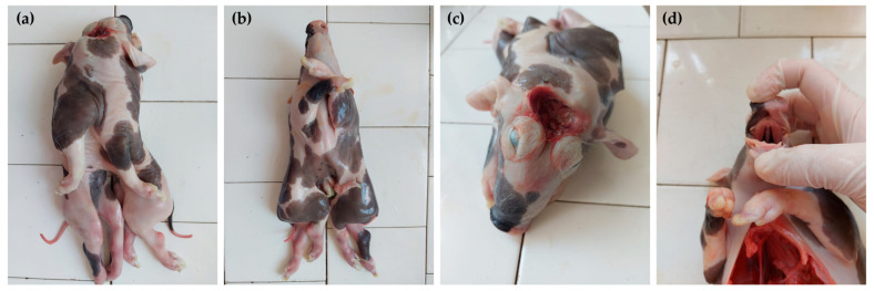
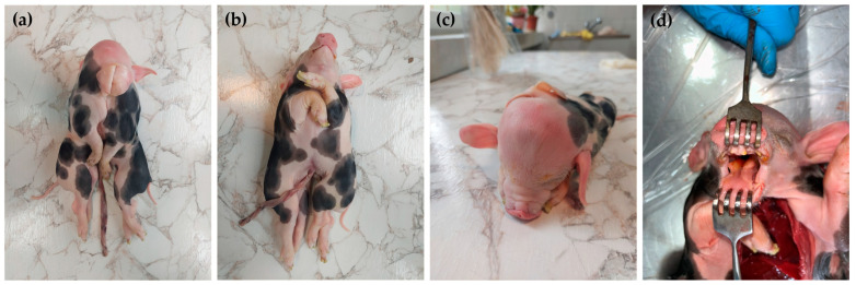
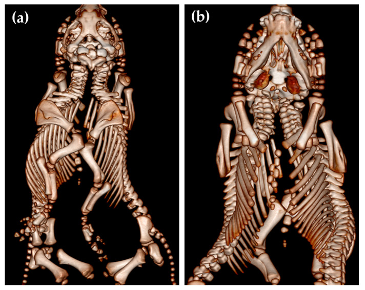
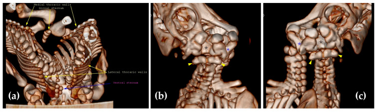
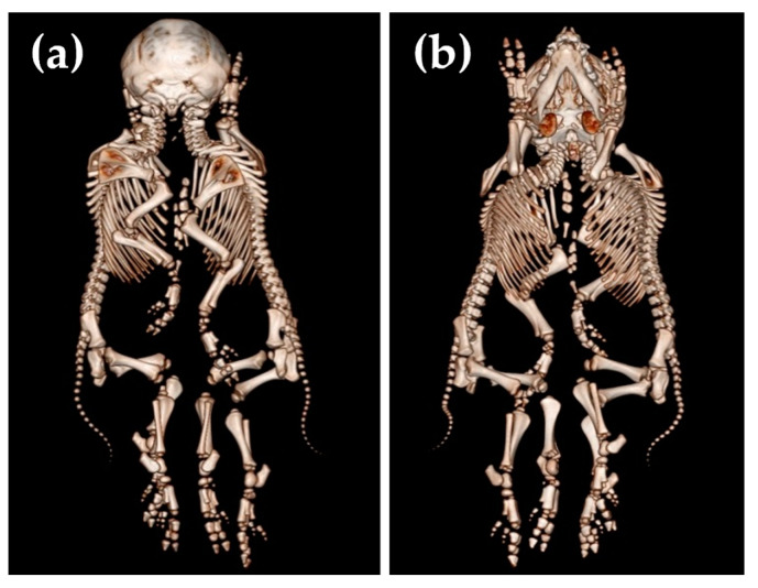
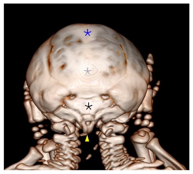
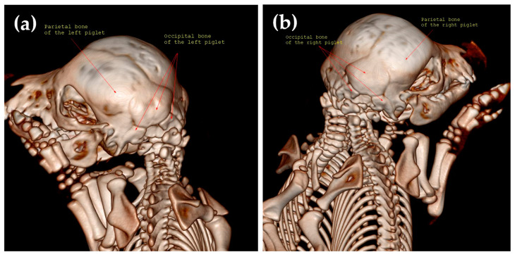
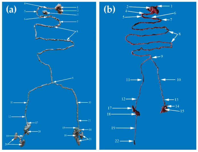
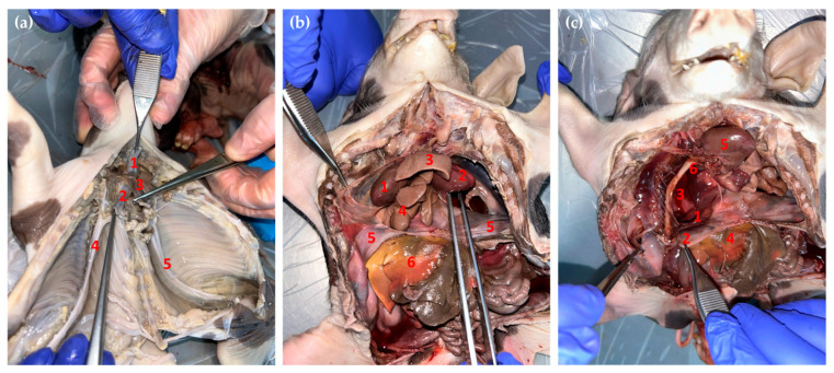
Conjoined twins are rare congenital malformations that have been reported in mammals. Two different cases are presented in this study. Case No. 1 features monocephalic, thoracopagus-conjoined twin piglets with anencephaly and palatoschisis of the Pietrain breed, and case No. 2 features monocephalic, thoracopagus conjoined twin piglets with palatoschisis and bifid root tongue of a mixed breed. These cases were examined using post-mortem and computed tomography (CT) examinations. In both cases, the conjoined symmetrical twins had a single head, one neck, and fused thoracic cavities, while the abdominal cavities were separated. Similarly, in both cases, they had four forelimbs and four hindlimbs and duplicated foramen magnum. During CT examination, in case No. 1, severe abnormalities were observed in the skull and vertebral column. In the left twin, occult dysraphism was seen from the C2 vertebra until the end of the vertebral column, and in the right twin, from the C3 vertebra until the end of the state vertebral level. In case No. 2, the oral cavity contained a tongue with a bifid root connected with one hyoid bone, and the soft palate presented a small cleft. During CT examination, the parietal bone and the occipital bones were partially duplicated. This case also presented occult dysraphism, but only in the cervical vertebrae, C1-C6 for the left twin and C1-C5 for the right twin. In both cases, abnormalities of the internal organs were revealed during necropsy. Conjoined twins with multiple congenital anomalies presented here enhance our understanding of the various clinical forms of conjoined cases in veterinary medicine.

Keywords: anencephaly; conjoined twins; embryogenesis; palatoschisis; swine congenital malformations.


Conflict of interest statement

The authors declare no conflicts of interest. The funders had no role in the design of the study, in the collection, analyses, or interpretation of the data, in the writing of the manuscript, or in the decision to publish the results.

Figures

Figure 1
Figure 1
The clinical aspects of the conjoined twins (case No. 1). Dorsal view (a), ventral view (b), rostral view of the head (c), and cleft palate view (d).
Figure 2
Figure 2
The clinical aspects of the conjoined twins (case No. 2). Dorsal view (a), ventral view (b), rostral view of the head (c), and bifid root tongue view (d).
Figure 3
Figure 3
Dorsal (a) and ventral (b) view of the conjoined twins (case No. 1).
Figure 4
Figure 4
CT scan of the thoracal cavities’ fusion (a). The right paracondylar process of the left twin fused with the left paracondylar process of the right twin (blue asterisk); there are two foramina magna (black asterisk), and the atlas is also indicated (yellow arrows). The squamous part of the occipital bone was duplicated and incompletely formed; a partial lack of parietal bone is seen in (b,c) (case No. 1).
Figure 5
Figure 5
Dorsal (a) and ventral (b) view of the conjoined twins, case No. 2.
Figure 6
Figure 6
Caudal view of the skull (case No. 2), where the blue asterisk represents the fusion between the right frontal bone of the left twin and the left frontal bone of the right twin, the grey asterisk represents the fusion between the right parietal bone of the left twin and the left parietal bone of the right twin, the black asterisk represents the fusion between the right temporal bone of the left twin and the left temporal bone of the right twin, and the yellow arrowhead represents the fusion between the right paracondylar process of the occipital bone of the left twin and the left paracondylar process of the occipital bone of the right twin.
Figure 7
Figure 7
The caudo-lateral view of the left twin (a) and the right twin (b).
Figure 8
Figure 8
The gastrointestinal tract of the conjoined twins, case No. 1 (a) and case No. 2 (b). 1. Esophagus; 2. Stomach; 3. Pancreas; 4. Descending duodenum; 5. Transverse duodenum; 6. Ascending duodenum; 7. Duodenojejunal flexure; 8. Jejunum; 9. Furcation of the jejunum into the left and right jejunum for the correspondent body; 10. Left terminal jejunum; 11. Right terminal jejunum; 12. Right ileum; 13. Left ileum; 14. Left cecum; 15. Left ascending colon; 16. Left descending colon; 17. Right cecum; 18. Right ascending colon; 19. Right descending colon; 20. Right kidneys; 21. Left kidneys; 22. Right rectum.
Figure 9
Figure 9
Necropsy of the conjoined twins. (a) Caudoventral aspect of the thoracic cavity (case No. 1): 1. Dorsal trachea; 2. Ventral trachea; 3. Esophagus; 4. Thoracic aorta from right body; 5. Thoracic aorta from left body. (b) Ventral view of the thoracic and abdominal cavities of the conjoined twins (case No. 2): 1. Right heart; 2. Left heart; 3. Lungs from the left body; 4. Lungs from the right body; 5. Diaphragm; 6. The liver of the right body. (c) Ventral view of the thoracic and abdominal cavities of the conjoined twins (case No. 2): 1. Diaphragmatic fissure; 2. Diaphragm; 3. The liver from the left body herniated into the thoracic cavities through the diaphragm; 4. Right liver; 5. Right heart; 6. Right thoracic aorta.

Similar articles

References

    1. Vanderzon D.M., Partlow G.D., Fisher K.R.S., Halina W.G. Parapagus conjoined twin holstein calf. Anat. Rec. 1998;251:60–65. doi: 10.1002/(SICI)1097-0185(199805)251:1<60::AID-AR10>3.0.CO;2-9. - DOI - PubMed
    1. Marzban Abbasabadi B., Ahmadzadeh A., Ramezanpour S., Hajati Ziabari A.R. A case of asymmetrical monocephalus dipygus (tetrapus dibrachius) in a male Holstein calf in Iran. Vet. Res. Forum. 2016;7:177–179. - PMC - PubMed
    1. Weber J., Behn H., Freick M. A rare case of monozygotic iniodymic diprosopiasis in a German Holstein calf. Tierärztliche Prax. Ausg. G Großtiere Nutztiere. 2017;45:296–301. doi: 10.15653/TPG-160724. - DOI - PubMed
    1. Kabak Y.B., Kabak M., Özak A., Sözmen M., İnal S., Güvenç T., Gülbahar M.Y. Asymmetric conjoined twins: Gnathopagus parasiticus. Ank. Üniversitesi Vet. Fakültesi Derg. 2020;67:431–435. doi: 10.33988/auvfd.618897. - DOI
    1. Prasad V.D., Kumar P.R., Krishna N.H., Sreenu M. Dystocia due to conjoined twins: A report of two cases. Int. J. Vet. Sci. Anim. Husb. 2016;1:28–29.

LinkOut - more resources