

Line-Field Confocal Optical Coherence Tomography for the Diagnosis of Skin Tumors: A Systematic Review and Meta-Analysis
- PMID: 39061659
- PMCID: PMC11276068
- DOI: 10.3390/diagnostics14141522
Line-Field Confocal Optical Coherence Tomography for the Diagnosis of Skin Tumors: A Systematic Review and Meta-Analysis
Abstract
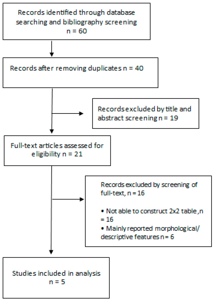
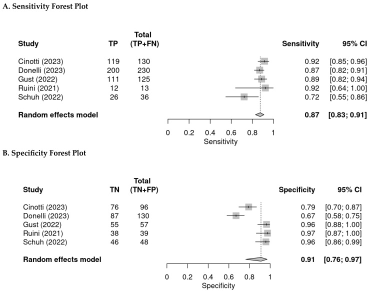
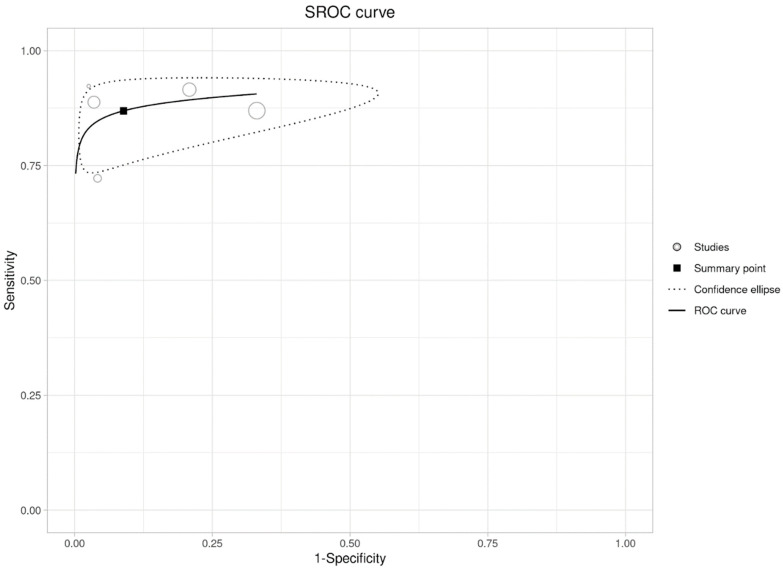
A line-field confocal optical coherence tomography (LC-OCT) combines confocal microscopy and optical coherence tomography into a single, rapid, easy-to-use device. This meta-analysis was performed to determine the reliability of LC-OCT for diagnosing malignant skin tumors. PubMed, EMBASE, Web of Science databases, and the Cochrane Library were searched for research studies in the English language from inception till December 2023. To assess quality and the risk of bias, the Quality Assessment of Diagnostic Accuracy Studies-2 (QUADAS-2) was used. The sensitivity and specificity of each study were calculated. The bivariate summary sensitivity and specificity were calculated using the linear mixed model. Five studies with 904 reported per lesion analyses in our study; the specificity and sensitivity ranged from 67% to 97% and 72% to 92%, respectively. The pooled specificity and sensitivity were 91% (95% CI: 76-97%) and 86.9% (95% CI: 81.8-90.8%), respectively. The summary sensitivity and specificity from the bivariate approach are 86.9% (95% CI: 81.8-90.8%) and 91.1% (95% CI: 76.7-97.0%), respectively. The area under the curve is 0.914. LC-OCT shows great sensitivity and specificity in diagnosing malignant skin tumors. However, due to the limited number of studies included in our meta-analysis, it is premature to elucidate the true potential of LC-OCT.
Keywords: basal cell carcinoma; line-field confocal optical coherence tomography; melanoma; squamous cell carcinoma.
Conflict of interest statement
Babar Rao is a consultant for Caliber ID (The manufacturer of VivaScope®). The other authors have no conflicts of interest to disclose.
Figures
Similar articles
-
Line-Field Confocal Optical Coherence Tomography for the Diagnosis of Skin Carcinomas: Real-Life Data over Three Years.Curr Oncol. 2023 Sep 28;30(10):8853-8864. doi: 10.3390/curroncol30100639. Curr Oncol. 2023. PMID: 37887539 Free PMC article.
-
Optical coherence tomography for the diagnosis of malignant skin tumors: a meta-analysis.J Biomed Opt. 2018 Feb;23(2):1-10. doi: 10.1117/1.JBO.23.2.020902. J Biomed Opt. 2018. PMID: 29473350
-
Line-Field Confocal Optical Coherence Tomography (LC-OCT) for Skin Imaging in Dermatology.Life (Basel). 2023 Nov 28;13(12):2268. doi: 10.3390/life13122268. Life (Basel). 2023. PMID: 38137869 Free PMC article.
-
Line-field confocal optical coherence tomography in melanocytic and non-melanocytic skin tumors.Ital J Dermatol Venerol. 2023 Jun;158(3):180-189. doi: 10.23736/S2784-8671.23.07639-9. Ital J Dermatol Venerol. 2023. PMID: 37278496 Review.
-
Line-field confocal optical coherence tomography-Practical applications in dermatology and comparison with established imaging methods.Skin Res Technol. 2021 May;27(3):340-352. doi: 10.1111/srt.12949. Epub 2020 Oct 21. Skin Res Technol. 2021. PMID: 33085784
Cited by
-
Image-Guided Radiation Therapy Is Equally Effective for Basal and Squamous Cell Carcinoma.Dermatopathology (Basel). 2024 Nov 19;11(4):315-329. doi: 10.3390/dermatopathology11040033. Dermatopathology (Basel). 2024. PMID: 39584849 Free PMC article.
-
Noninvasive Multimodal Imaging and Its Role in Diagnosing Skin Lesions in Dermatology: A Systematic Review and Meta-Analysis.Am J Clin Dermatol. 2025 Jul 8. doi: 10.1007/s40257-025-00958-4. Online ahead of print. Am J Clin Dermatol. 2025. PMID: 40627274
-
Handheld multiphoton and pinhole-free reflectance confocal microscopy enables noninvasive, real-time cross-sectional imaging in skin.Sci Rep. 2024 Oct 30;14(1):26129. doi: 10.1038/s41598-024-76908-7. Sci Rep. 2024. PMID: 39478114 Free PMC article.
-
In Vivo Reflectance Confocal Microscopy Applied to Acral Melanocytic Lesions: A Systematic Review of the Literature.Diagnostics (Basel). 2024 Sep 25;14(19):2134. doi: 10.3390/diagnostics14192134. Diagnostics (Basel). 2024. PMID: 39410538 Free PMC article. Review.
References
-
- Donelli C., Suppa M., Tognetti L., Perrot J.L., Calabrese L., Pérez-Anker J., Malvehy J., Rubegni P., Cinotti E. Line-Field Confocal Optical Coherence Tomography for the Diagnosis of Skin Carcinomas: Real-Life Data over Three Years. Curr. Oncol. 2023;30:8853–8864. doi: 10.3390/curroncol30100639. - DOI - PMC - PubMed
-
- Schwartz M., Levine A., Markowitz O. Optical coherence tomography in dermatology. Cutis. 2017;100:163–166. - PubMed
Publication types
LinkOut - more resources
Full Text Sources
