

Adverse Effects Associated with Dermal Filler Treatments: Part II Vascular Complication
- PMID: 39061692
- PMCID: PMC11276034
- DOI: 10.3390/diagnostics14141555
Adverse Effects Associated with Dermal Filler Treatments: Part II Vascular Complication
Abstract
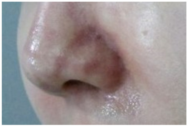
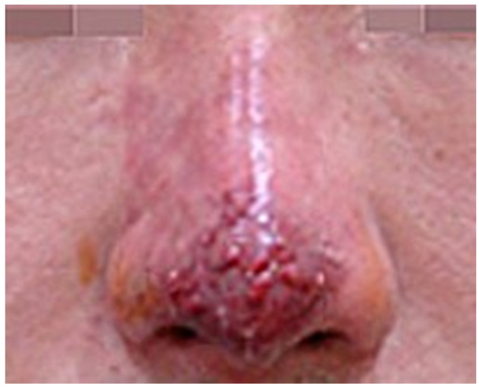
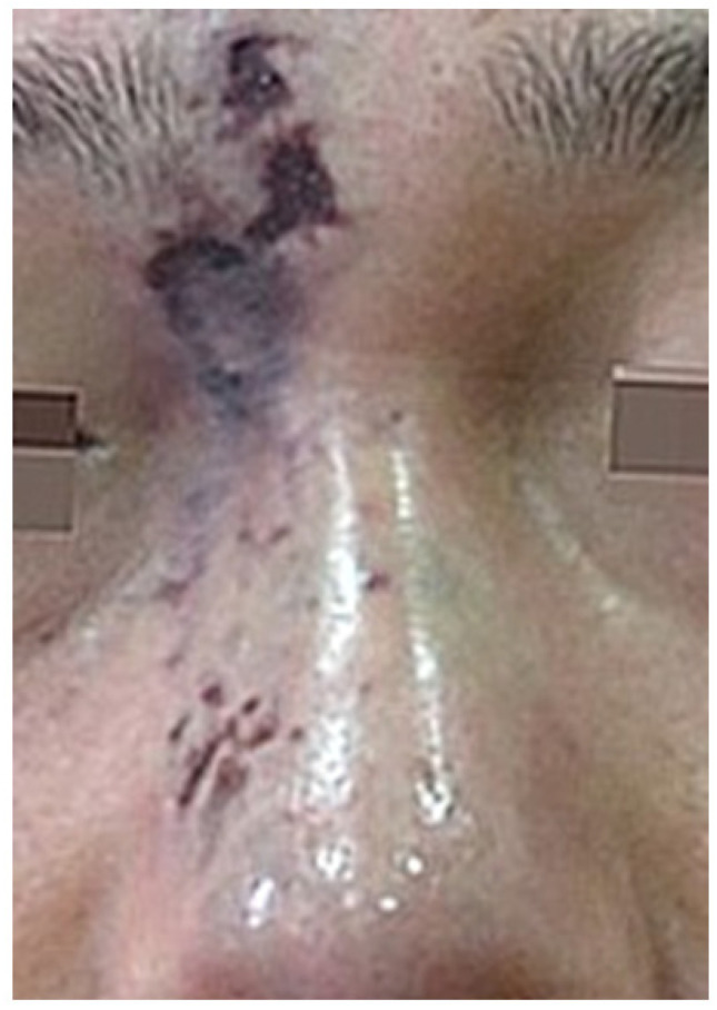
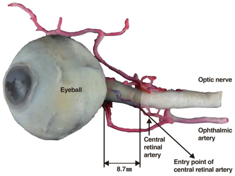
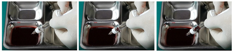
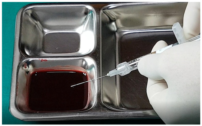
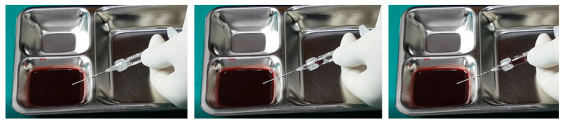
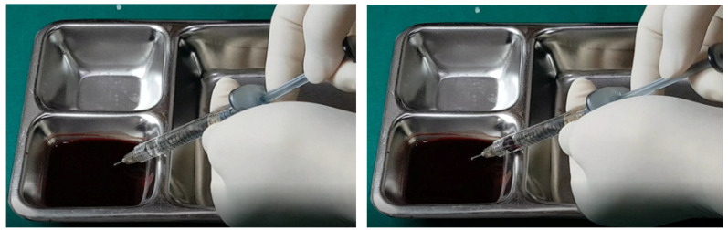
Vascular complications arising from dermal filler treatments pose significant risks, including ischemia, tissue necrosis, and severe outcomes like blindness and pulmonary embolism. This study investigates the mechanisms of vascular complications, categorizing them into extravascular compression and intravascular emboli. Extravascular compression occurs when injected fillers compress adjacent blood vessels, leading to ischemia and potential necrosis, while intravascular emboli result from fillers entering blood vessels, causing blockages. The study emphasizes the importance of anatomical knowledge, careful injection techniques, and early intervention. Management strategies include the use of hyaluronidase to dissolve HA fillers, vasodilators to improve blood circulation, and hyperbaric oxygen therapy. The regions most susceptible to complications align with major arterial pathways, particularly the nasolabial folds and nasal region. The study also highlights the need for meticulous injection techniques, the use of cannulas over needles in high-risk areas, and the aspiration test to detect vessel penetration. Early detection and immediate intervention are crucial to mitigate adverse outcomes. Continuous education and training for practitioners, along with advancements in filler materials and injection methods, are essential for improving the safety of cosmetic procedures. This comprehensive understanding aids in preventing and managing vascular complications, ensuring better patient outcomes. The field of dermal filler treatments is advancing with new techniques and technologies, such as High-Resolution Ultrasound, Infrared Imaging, self-crossing hyaluronic acid filler, biodegradable microspheres, and microinjection.
Keywords: blindness; cannulas; dermal fillers; hyaluronic acid; hyperbaric oxygen therapy; injection techniques; ischemia; pulmonary embolism; tissue necrosis; vascular complications.
Conflict of interest statement
I acknowledge that I have considered the conflict of interest statement included in the “Author Guidelines”. I hereby certify that, to the best of my knowledge, no aspect of my current personal or professional situation might reasonably be expected to significantly affect my views on the subject I am presenting.
Figures
References
-
- Sorensen E.P., Urman C. Cosmetic complications: Rare and serious events following botulinum toxin and soft tissue filler administration. J. Drugs Dermatol. 2015;14:486–491. - PubMed
Publication types
LinkOut - more resources
Full Text Sources
