. 2024 Jun 21;11(7):637.
doi: 10.3390/bioengineering11070637.

Synergistic Cellular Responses Conferred by Concurrent Optical and Magnetic Stimulation Are Attenuated by Simultaneous Exposure to Streptomycin: An Antibiotic Dilemma

Affiliations

Synergistic Cellular Responses Conferred by Concurrent Optical and Magnetic Stimulation Are Attenuated by Simultaneous Exposure to Streptomycin: An Antibiotic Dilemma

Jan Nikolas Iversen et al. Bioengineering (Basel). .

Abstract

Concurrent optical and magnetic stimulation (COMS) combines extremely low-frequency electromagnetic and light exposure for enhanced wound healing. We investigated the potential mechanistic synergism between the magnetic and light components of COMS by comparing their individual and combined cellular responses. Lone magnetic field exposure produced greater enhancements in cell proliferation than light alone, yet the combined effects of magnetic fields and light were supra-additive of the individual responses. Reactive oxygen species were incrementally reduced by exposure to light, magnetics fields, and their combination, wherein statistical significance was only achieved by the combined COMS modality. By contrast, ATP production was most greatly enhanced by magnetic exposure in combination with light, indicating that mitochondrial respiratory efficiency was improved by the combination of magnetic fields plus light. Protein expression pertaining to cell proliferation was preferentially enhanced by the COMS modality, as were the protein levels of the TRPC1 cation channel that had been previously implicated as part of a calcium-mitochondrial signaling axis invoked by electromagnetic exposure and necessary for proliferation. These results indicate that light facilitates functional synergism with magnetic fields that ultimately impinge on mitochondria-dependent developmental responses. Aminoglycoside antibiotics (AGAs) have been previously shown to inhibit TRPC1-mediated magnetotransduction, whereas their influence over photomodulation has not been explored. Streptomycin applied during exposure to light, magnetic fields, or COMS reduced their respective proliferation enhancements, whereas streptomycin added after the exposure did not. Magnetic field exposure and the COMS modality were capable of partially overcoming the antagonism of proliferation produced by streptomycin treatment, whereas light alone was not. The antagonism of photon-electromagnetic effects by streptomycin implicates TRPC1-mediated calcium entry in both magnetotransduction and photomodulation. Avoiding the prophylactic use of AGAs during COMS therapy will be crucial for maintaining clinical efficacy and is a common concern in most other electromagnetic regenerative paradigms.

Keywords: aminoglycoside antibiotics; chronic wounds; hard-to-heal wounds; magnetic mitohormesis; magnetoreception; mitochondria; photomodulation; proliferation; reactive oxygen species; wound healing.


Conflict of interest statement

J.F. is a cofounder of Piomic Medical AG. J.F. is also the founder of Fields at Work GmbH, a company that specializes in designing and manufacturing magnetic radiation exposure and detection devices. Fields at Work GmbH operates independently of Piomic Medical AG. All other authors declare no conflicts of interest.

Figures

Figure 1
Figure 1
Incremental myoblast proliferation induction upon exposure to light, magnetic fields, or their combination in the absence of streptomycin. (A) Live cell count of murine C2C12 myoblasts in response to sham (black), light (red), magnetic fields (blue), or combined COMS (hatched blue/red) exposure. (B) Table of fold changes in live cell count relative to the sham condition. The gray shaded area represents the absence of streptomycin at all times during exposure to the indicated conditions. Statistical analyses were performed minimally in three independent biological replicates. Data were analyzed using one-way ANOVA followed by multiple comparison tests. Significance levels are indicated as follows: * p < 0.05, ** p < 0.01, *** p < 0.001, and **** p < 0.0001. Error bars represent the standard error of the mean (SEM).
Figure 2
Figure 2
The timing of streptomycin administration produced differential effects over myogenic proliferation induction in response to light and magnetic field combinations. (A) Schematic illustration of when streptomycin (100 µg/mL) was added to the bathing media of cells in the “Strep During” (blue shaded) and “Strep After” (red shaded) paradigms prior to cell enumeration at 24 h after the indicated interventions. (B) Live cell count of mouse murine myoblasts in response to the different stimuli as aforementioned with streptomycin antibiotic supplementation (100 µg/mL) added 15 min before (left side, blue shaded box) or after (right side, red shaded box) exposure to the intervention. (C) Table showing the fold change of live cell count over sham condition (without streptomycin administration). The shaded areas indicate the absence of streptomycin (gray) or its application before and during (blue) or after exposure (red) to the indicated conditions. All data collected were from cells of the same plating. Statistical analyses were performed minimally in three independent biological replicates. Data were analyzed using one-way ANOVA followed by multiple comparison tests. Significance levels are indicated as follows: ** p < 0.01, and **** p < 0.0001. The error bars represent the standard error of the mean (SEM).
Figure 3
Figure 3
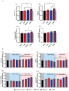
Myogenic proliferation associated protein expression in response to exposure to light, magnetic fields, and their combination. (A) Protein expression of (i) cyclin D1, (ii) TRPC1, (iii) phosphorylated ERK, (iv) and cyclin B1 in response to the indicated exposure intervention (n = 3) in the absence of streptomycin. (B) Protein expressions of (i) cyclin D1, (ii) TRPC1, (iii) phosphorylated ERK, (iv) and cyclin B1 either in the presence of streptomycin (100 µg/mL) during (blue shaded box) or after (red shaded box) exposure as indicated (n = 3). The shaded areas indicate the absence of streptomycin (gray) or its application before and during (blue) or after exposure (red) to the indicated conditions. All data shown in panel (B) were collected from cells of the same plating and represent independent cell samples as those used in panel (A). Statistical analyses were performed minimally in three independent biological replicates. Data were analyzed using one-way ANOVA followed by multiple comparison tests. Significance levels are indicated as follows: * p < 0.05 and ** p < 0.01. The error bars represent the standard error of the mean (SEM).
Figure 4
Figure 4
Dichotomous changes in ROS and ATP production following COMS exposure. (A) Bar chart showing the immediate ROS response of cells (fluorescent intensity averaged during initial 3 min of reading) for the indicated interventions (n = 4). (B) ROS level of cells expressed as fluorescent intensity fold change at 17 min (t17) over time 0, (t0), (n = 5). (C) The bar chart shows the ATP levels of cells (expressed as fold change over Sham) at t17 (n = 5, with six technical replicates each). In all cases, cells were exposed for 5 min to the indicated exposure modality at the start of device activation before measurements were commenced. The presented data were generated in the absence of streptomycin. Data were analyzed using one-way ANOVA followed by multiple comparison tests with * p < 0.05 and *** p < 0.001. The error bars represent the standard error of the mean (SEM).

Similar articles

Cited by

References

    1. Reinboldt-Jockenhofer F., Traber J., Liesch G., Bittner C., Benecke U., Dissemond J. Concurrent optical and magnetic stimulation therapy in patients with lower extremity hard-to-heal wounds. J. Wound Care. 2022;31:S12–S21. doi: 10.12968/jowc.2022.31.Sup6.S12. - DOI - PubMed
    1. Traber J., Wild T., Marotz J., Berli M.C., Franco-Obregon A. Concurrent Optical- and Magnetic-Stimulation-Induced Changes on Wound Healing Parameters, Analyzed by Hyperspectral Imaging: An Exploratory Case Series. Bioengineering. 2023;10:750. doi: 10.3390/bioengineering10070750. - DOI - PMC - PubMed
    1. Franco-Obregon A. Harmonizing Magnetic Mitohormetic Regenerative Strategies: Developmental Implications of a Calcium-Mitochondrial Axis Invoked by Magnetic Field Exposure. Bioengineering. 2023;10:1176. doi: 10.3390/bioengineering10101176. - DOI - PMC - PubMed
    1. Yap J.L.Y., Tai Y.K., Fröhlich J., Fong C.H.H., Yin J.N., Foo Z.L., Ramanan S., Beyer C., Toh S.J., Casarosa M., et al. Ambient and supplemental magnetic fields promote myogenesis via a TRPC1-mitochondrial axis: Evidence of a magnetic mitohormetic mechanism. FASEB J. 2019;33:12853–12872. doi: 10.1096/fj.201900057R. - DOI - PMC - PubMed
    1. Ma Q., Chen C., Deng P., Zhu G., Lin M., Zhang L., Xu S., He M., Lu Y., Duan W., et al. Extremely Low-Frequency Electromagnetic Fields Promote In Vitro Neuronal Differentiation and Neurite Outgrowth of Embryonic Neural Stem Cells via Up-Regulating TRPC1. PLoS ONE. 2016;11:e0150923. doi: 10.1371/journal.pone.0150923. - DOI - PMC - PubMed

LinkOut - more resources