

One-Step Intraoperative Optical Coherence Tomography Guided Tunnel, Mushroom Femtosecond Laser Big Bubble Deep Anterior Lamellar Keratoplasty
- PMID: 39061721
- PMCID: PMC11273850
- DOI: 10.3390/bioengineering11070639
One-Step Intraoperative Optical Coherence Tomography Guided Tunnel, Mushroom Femtosecond Laser Big Bubble Deep Anterior Lamellar Keratoplasty
Abstract
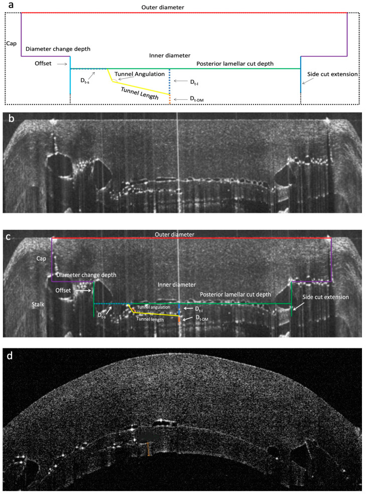
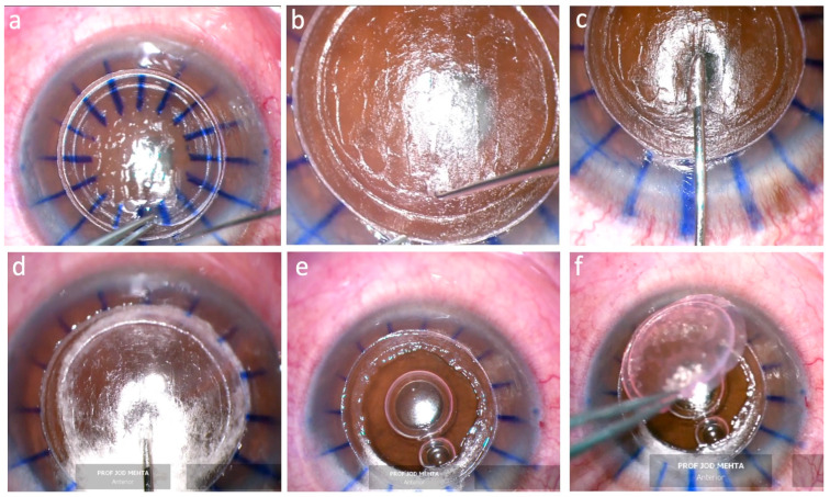
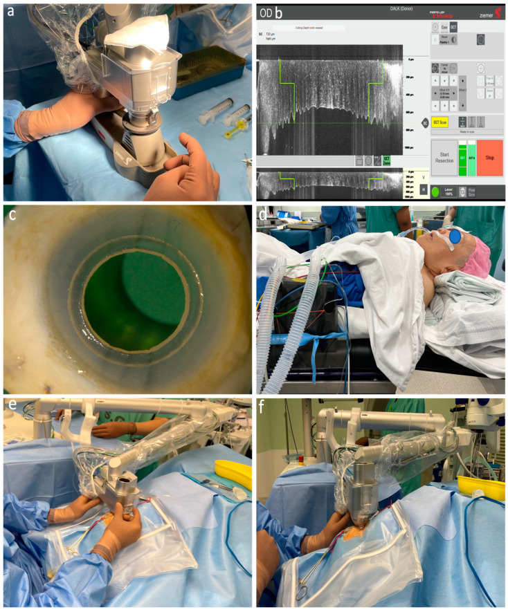
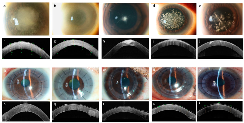
The aim of our study is to investigate the feasibility and outcomes of using a femtosecond laser (FSL) platform (Ziemer LDV Z8) for deep anterior lamellar keratoplasty (DALK), enabling the creation of mushroom-shaped graft-host junctions, lamellar cuts, and intrastromal tunnels, to facilitate the big bubble, in one step. We included wet lab experiments on nine porcine eyes to assess the laser accuracy and cuts depth using an anterior segment (AS) OCT. This was followed by an interventional prospective case series on 10 eyes with variant corneal pathologies. The Z8 system, with in-built intraoperative optical coherence tomography (iOCT), guided corneal scans and directed the cuts. ASOCT showed visible mushroom configurations, lamellar cuts, and tunnels. Deviations from the target were 1.6%, 2.6%, and 3.5%. Anterior lamellar removal was easy in all clinical cases, including corneal scarring. The intrastromal tunnel was found at the preset location and the mushroom configuration was acquired. A big bubble was achieved in all cases. Type 1, 2, and 3 bubbles were formed in eight, one, and one case, respectively. We describe a new approach to DALK in which the in-built iOCT-guided FSL enables safe, precise, controlled, and reproducible desired cuts in one step. The preliminary clinical outcomes were favorable.
Keywords: deep anterior lamellar keratoplasty; femtosecond laser; intraoperative optical coherent tomography; intrastromal tunnel; mushroom configuration.
Conflict of interest statement
The authors declare no conflict of interest.
Figures
Similar articles
-
Intraoperative Optical Coherence Tomography-Guided Femtosecond Laser-Assisted Deep Anterior Lamellar Keratoplasty.Cornea. 2019 May;38(5):648-653. doi: 10.1097/ICO.0000000000001851. Cornea. 2019. PMID: 30614905
-
A novel method of tunnel creation using intraoperative optical coherence tomography-guided deep anterior lamellar keratoplasty.Indian J Ophthalmol. 2021 Dec;69(12):3743-3744. doi: 10.4103/ijo.IJO_531_21. Indian J Ophthalmol. 2021. PMID: 34827035 Free PMC article.
-
Clinical comparison of manual and laser-cut corneal tunnel for intrastromal air injection in femtosecond laser-assisted deep anterior lamellar keratoplasty (DALK).Graefes Arch Clin Exp Ophthalmol. 2023 Jan;261(1):185-191. doi: 10.1007/s00417-022-05765-9. Epub 2022 Jul 28. Graefes Arch Clin Exp Ophthalmol. 2023. PMID: 35896678
-
An Overview of Intraoperative OCT-Assisted Lamellar Corneal Transplants: A Game Changer?Diagnostics (Basel). 2022 Mar 17;12(3):727. doi: 10.3390/diagnostics12030727. Diagnostics (Basel). 2022. PMID: 35328280 Free PMC article. Review.
-
[Intraoperative Optical Coherence Tomography In Deep Anterior Lamellar Keratoplasty].Klin Monbl Augenheilkd. 2016 Jun;233(6):717-21. doi: 10.1055/s-0042-108588. Epub 2016 Jun 17. Klin Monbl Augenheilkd. 2016. PMID: 27315292 Review. German.
Cited by
-
Intraoperative Optical Coherence Tomography (OCT)-Guided Femtosecond Laser-Assisted Descemet Membrane Endothelial Keratoplasty (iFAD).Bioengineering (Basel). 2024 Nov 25;11(12):1192. doi: 10.3390/bioengineering11121192. Bioengineering (Basel). 2024. PMID: 39768010 Free PMC article.
References
LinkOut - more resources
Full Text Sources
Research Materials
Miscellaneous
