. 2024 Jul 15;11(7):718.
doi: 10.3390/bioengineering11070718.

CARING: Cannula for Alleviation of Retinal Injury Caused by Needle Fluidic Gashing

Affiliations

CARING: Cannula for Alleviation of Retinal Injury Caused by Needle Fluidic Gashing

Kaersti L Rickels et al. Bioengineering (Basel). .

Abstract

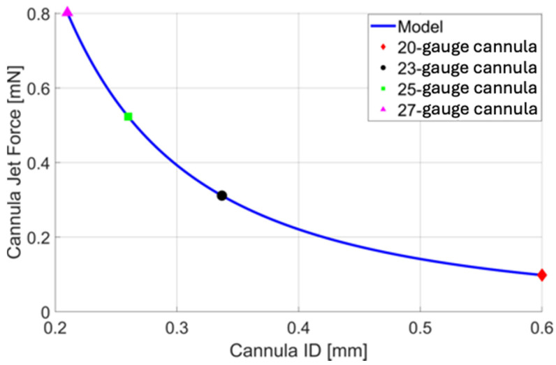
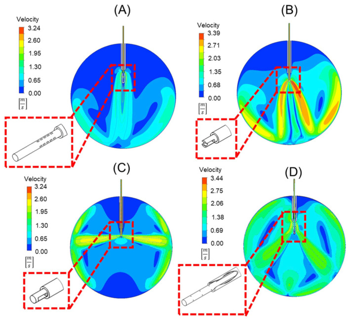
Infusion-related iatrogenic retinal breaks (IRBs) are a significant complication in vitrectomies, particularly when smaller-gauge cannulas are used during fluid infusion. Using two-dimensional finite element analysis (FEA), we analyzed forces exerted on the retina from different cannulas: traditional 25-gauge, 20-gauge, 23-gauge, and 27-gauge, then investigated four alternative new cannula designs: (A) oblique orifices, (B) external obstruction, (C) side ports, and (D) perpendicular orifices. The analysis revealed that the standard 25-gauge cannula had a force of 0.546 milli-Newtons (mN). Optimized cannulas demonstrated decreased forces: 0.072 mN (A), 0.266 mN (B), 0.417 mN (C), and 0.117 mN (D). While all the designs decrease fluid jet force, each has unique challenges: Design A may complicate manufacturing, B requires unique attachment techniques, C could misdirect fluid toward the lens and peripheral retina, and D requires a sealed trocar/cannula design to prevent unwanted fluid ejection. These four innovative cannula designs, identified with detailed engineering simulations, provide promising strategies to reduce the risk of IRBs during vitrectomy, bridging the gap between engineering insights and clinical application.

Keywords: cannula tip design; fluidic gashing; iatrogenic retinal breaks; infusion breaks.


Conflict of interest statement

The authors declare no conflict of interest.

Figures

Figure 1
Figure 1
(A) FEA result of the water volume fraction using a 20-gauge cannula system with a 24 mm eye. In the detailed view (inset), the width parameter w (white arrows) can be seen depicted. The water jet has a velocity u (red), that exerts a distributed force, w (four black arrows). This distributed force produces a total effective force F (single black arrow). Blue represents 0% water (i.e., air) and red represents 100% water. (B) FEA result of the water volume fraction with orifices perpendicular to the longitudinal axis of the cannula.
Figure 2
Figure 2
(A) Mesh resulting from a cell size of 1e-4 m. (B) Resulting volume fraction contour after initialization and patch (blue represents air in this figure).
Figure 3
Figure 3
A plot of cannula infusion fluid stream force vs. cannula inner diameter (ID) based on Equation 5 with a volumetric flow rate of 10 mLmin. For quick reference, the force resulting from the infusion fluid stream of a 20-gauge (red), 23-gauge (black), 25-gauge (green), and 27-gauge (magenta) cannula is presented, allowing us to assess their nominal cannula designs.
Figure 4
Figure 4
A comparison of a 20-gauge cannula (A,B) to a 25-gauge cannula (C,D). The water volume fraction contours (A,C) can be seen alongside the velocity contours (B,D). Note that the scales are not the same for the velocity plot, with red representing 0.78 [m/s] in (B) and 3.41 [m/s] in (D).
Figure 5
Figure 5
Velocity contours of four different cannula designs. These include (A) oblique orifices at an angle of 60°, (B) an external obstruction similar to a spark plug, (C) orifices perpendicular to the longitudinal axis of the cannula, and (D) a design that uses orifices in the cannula within, and outside of, the trocar body to disperse flow. Depicted in each is a detailed view of the geometry of the trocar and cannula in 3-D.

Similar articles

References

    1. Lalezary M., Kim S.J., Jiramongkolchai K., Recchia F.M., Agarwal A., Sternberg P., Jr. Long-Term Trends in Intraocular Pressure after Pars Plana Vitrectomy. Retina. 2011;31:679–685. doi: 10.1097/IAE.0b013e3181ff0d5a. - DOI - PubMed
    1. Scott M.N., Weng C.Y. The Evolution of Pars Plana Vitrectomy to 27-G Microincision Vitrectomy Surgery. Int. Ophthalmol. Clin. 2016;56:97–111. doi: 10.1097/IIO.0000000000000131. - DOI - PubMed
    1. Mohamed S., Claes C., Tsang C.W. Review of Small Gauge Vitrectomy: Progress and Innovations. J. Ophthalmol. 2017;2017:6285869. doi: 10.1155/2017/6285869. - DOI - PMC - PubMed
    1. Omari A., Mahmoud T.H. Vitrectomy. StatPearls Publishing; Treasure Island, FL, USA: 2020. - PubMed
    1. Chen E. 25-Gauge Transconjunctival Sutureless Vitrectomy. Curr. Opin. Ophthalmol. 2007;18:188–193. doi: 10.1097/ICU.0b013e328133889a. - DOI - PubMed

LinkOut - more resources