. 2024 Jul 16;11(7):721.
doi: 10.3390/bioengineering11070721.

Developing a Swallow-State Monitoring System Using Nasal Airflow, Surface Electromyography, and Thyroid Cartilage Movement Detection

Affiliations

Developing a Swallow-State Monitoring System Using Nasal Airflow, Surface Electromyography, and Thyroid Cartilage Movement Detection

Wann-Yun Shieh et al. Bioengineering (Basel). .

Abstract

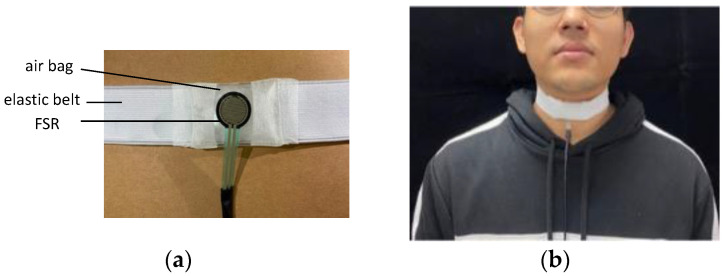
The safe ingestion of food and water requires appropriate coordination between the respiratory and swallowing pathways. This coordination can be disrupted because of aging or various diseases, thereby resulting in swallowing disorders. No comparative research has been conducted on methods for effectively screening swallowing disorders in individuals and providing timely alerts to their caregivers. Therefore, the present study developed a monitoring and alert system for swallowing disorders by using three types of noninvasive sensors, namely those measuring nasal airflow, surface electromyography signals, and thyroid cartilage movement. Two groups of participants, one comprising healthy individuals (58 participants; mean age 49.4 years) and another consisting of individuals with a history of unilateral stroke (21 participants; mean age 54.4 years), were monitored when they swallowed five volumes of water. Through an analysis of the data from both groups, seven indicators of swallowing disorders were identified, and the proposed system characterized the individual's swallowing state as having a green (safe), yellow (unsafe), or red (highly unsafe) status on the basis of these indicators. The results indicated that the symptoms of swallowing disorders are detectable. Healthcare professionals can then use these data to conduct assessments, perform screening, and provide nutrient intake suggestions.

Keywords: noninvasive sensor; respiration and swallow coordination; swallow state monitoring; swallowing disorder.


Conflict of interest statement

The authors declare no conflicts of interest.

Figures

Figure 1
Figure 1
Proposed alert method for swallowing disorders.
Figure 2
Figure 2
Surface electromyography (sEMG) measurement: (a) bipolar electrodes and (b) attachment of the sEMG electrodes to the human body.
Figure 3
Figure 3
Nasal airflow measurement: (a) cannula and pressure transducer and (b) placement of the nasal cannula.
Figure 4
Figure 4
Measurement with the FSR: (a) FSR with an elastic belt and (b) placement of the FSR belt.
Figure 5
Figure 5
Design of the adopted signal collector, including the top view and side view.
Figure 6
Figure 6
Swallowing and respiration signals acquired by the three types of adopted sensors. In this figure, E1–E3, A1 to A2, F1 to F2, and F2 to F3 represent durations of the (1) submental muscle response, (2) respiration apnea, (3) upward and forward movement of the thyroid cartilage to block the trachea, and (4) movement of the thyroid cartilage back to its original position, respectively.
Figure 7
Figure 7
Three-color alert model for the swallowing state. Green, yellow, and red correspond to a normal state, Level 2 alerts, and Level 1 alerts, respectively.
Figure 8
Figure 8
Mean total excursion time (TET) values for the healthy and patient groups.
Figure 9
Figure 9
Mean durations of respiration apnea (SAD) for the patient and healthy groups.
Figure 10
Figure 10
Mean onset latency between sEMG signals and thyroid cartilage excursion (OL) for the two groups.
Figure 11
Figure 11
Mean durations of the second deflection in the W-shaped response of the FSR (D2DEF) for the two groups.
Figure 12
Figure 12
Mean durations of sEMG signals for submentalis activity (DsEMG) for the two groups.

Similar articles

Cited by

References

    1. Barczi S.R., Sullivan P.A., Robbins J.A. How should dysphagia care of older adults differ? Establishing optimal practice patterns. Semin. Speech Lang. 2000;21:0347–0364. doi: 10.1055/s-2000-8387. - DOI - PubMed
    1. Cichero J.A. Age-related changes to eating and swallowing impact frailty: Aspiration, choking risk, modified food texture and autonomy of choice. Geriatrics. 2018;3:69. doi: 10.3390/geriatrics3040069. - DOI - PMC - PubMed
    1. Blumberg R., Feldman C., Murray D., Burnes N., Murawski D. Food and nutrition care in long-term care facilities: Examining the perspectives of frontline workers. J. Nutr. Gerontol. Geriatr. 2018;37:145–157. doi: 10.1080/21551197.2018.1516593. - DOI - PubMed
    1. Ertekin C. Physiological and pathological aspects of oropharyngeal swallowing. Mov. Disord. 2002;17:S86–S89. doi: 10.1002/mds.10068. - DOI - PubMed
    1. Boaden E., Nightingale J., Bradbury C., Hives L., Georgiou R. Clinical practice guidelines for videofluoroscopic swallowing studies: A systematic review. Radiography. 2020;26:154–162. doi: 10.1016/j.radi.2019.10.011. - DOI - PubMed

LinkOut - more resources