Review
. 2024 Jun 26;13(7):768.
doi: 10.3390/antiox13070768.

Sodium-Glucose Cotransporter Inhibitors: Cellular Mechanisms Involved in the Lipid Metabolism and the Treatment of Chronic Kidney Disease Associated with Metabolic Syndrome

Affiliations
Review

Sodium-Glucose Cotransporter Inhibitors: Cellular Mechanisms Involved in the Lipid Metabolism and the Treatment of Chronic Kidney Disease Associated with Metabolic Syndrome

Fernando Cortés-Camacho et al. Antioxidants (Basel). .

Abstract

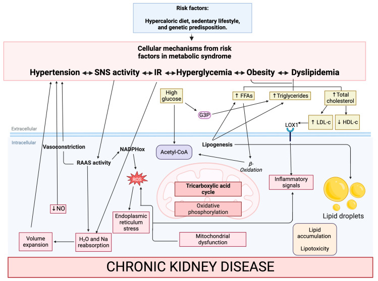
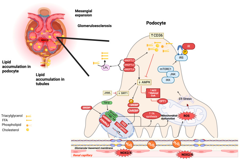
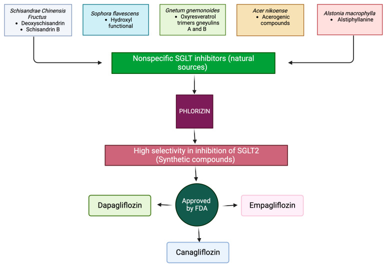
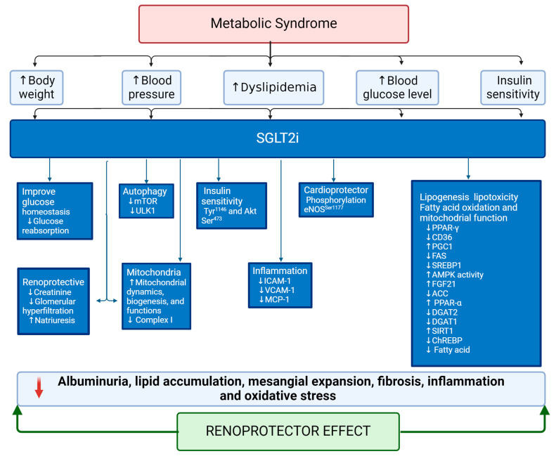
Metabolic syndrome (MetS) is a multifactorial condition that significantly increases the risk of cardiovascular disease and chronic kidney disease (CKD). Recent studies have emphasized the role of lipid dysregulation in activating cellular mechanisms that contribute to CKD progression in the context of MetS. Sodium-glucose cotransporter 2 inhibitors (SGLT2i) have demonstrated efficacy in improving various components of MetS, including obesity, dyslipidemia, and insulin resistance. While SGLT2i have shown cardioprotective benefits, the underlying cellular mechanisms in MetS and CKD remain poorly studied. Therefore, this review aims to elucidate the cellular mechanisms by which SGLT2i modulate lipid metabolism and their impact on insulin resistance, mitochondrial dysfunction, oxidative stress, and CKD progression. We also explore the potential benefits of combining SGLT2i with other antidiabetic drugs. By examining the beneficial effects, molecular targets, and cytoprotective mechanisms of both natural and synthetic SGLT2i, this review provides a comprehensive understanding of their therapeutic potential in managing MetS-induced CKD. The information presented here highlights the significance of SGLT2i in addressing the complex interplay between metabolic dysregulation, lipid metabolism dysfunction, and renal impairment, offering clinicians and researchers a valuable resource for developing improved treatment strategies and personalized approaches for patients with MetS and CKD.

Keywords: chronic kidney disease; dyslipidemia; hypertension; insulin resistance; lipid metabolism; lipotoxicity; metabolic syndrome; obesity; oxidative stress; sodium–glucose cotransporter 2 inhibitors.


Conflict of interest statement

The authors declare no conflicts of interest.

Figures

Figure 1
Figure 1
Cellular mechanisms from risk factors in metabolic syndrome and their role in kidney damage. Metabolic dysfunction alters the lipid profile, leading to the activation of inflammatory processes, oxidative stress, and increased lipid accumulation in the circulation and renal tissue. Insulin resistance and dyslipidemia deteriorate mitochondrial function, favoring ROS production and cell apoptosis. On the other hand, SNS hyperactivity promotes renal sodium reabsorption, leading to hypertension and renal injury. Additionally, the high activity of RAAS contributes to ROS and affects renal hemodynamics, sodium retention, and vasoconstriction, which causes kidney damage. Abbreviations: FFAs, free fatty acids; G3P, glyceraldehyde 3-phosphate; HDL-c, high-density lipoprotein cholesterol; LDL-c, low-density lipoprotein cholesterol; SNS, sympathetic nervous system; ROS, reactive oxygen species; RAAS, renin–angiotensin–aldosterone system; LOX1, lectin-like oxidized low-density lipoprotein receptor-1; NO, nitric oxide. IR, insulin resistance; NADPHox, nicotinamide adenine dinucleotide phosphate oxidase.
Figure 2
Figure 2
Role of lipids in chronic kidney disease. CD36 is responsible for transporting FFA in podocytes, and these are stored in lipid drops, which inhibits AMPK activity, increasing the levels of ACC and malonyl-CoA and inactivating the CPT1 enzyme important for FAS; under these conditions, lipid synthesis and lipotoxicity increase. SREBP1 and ChREBP proteins can induce overexpression of lipogenic genes that favor de novo lipogenesis. Overexpression of JAML in podocytes affects lipid accumulation via SIRT1-AMPK-SREBP1, causing renal dysfunction. ANGPTL3, ANGPTL4, and ANGPTL8 are key regulators of LPL, which can reduce triglyceride levels to FFA, resulting in hypertriglyceridemia and causing damage to the structure of podocytes and proteinuria. Alternately, TGF-β decreases the expression of PGC-1α and, through Smad3, activates the synthesis of fatty acids. Low expression of PPARα exhibits higher lipid accumulation, while elevated levels of PPARγ increase lipogenesis. On the other hand, excess cholesterol increases the production of ROS by NOX4/NOX2, increasing H2O2. OS from high concentrations of fatty acids in podocytes induces IR by activating PKC; NF-κB; and JNK. In addition, several kinases, such as IKKβ; ERK, and mTORC1, are activated, resulting in phosphorylation of IRS-1 at inhibitory sites leading to IR. The black arrow indicates stimulation and the red lines indicate inhibition. Abbreviations: ANGPTL3, angiopoietin-like 3; ANGPTL4, angiopoietin-like 4; ANGPTL8, angiopoietin-like 8; CD36, cluster of differentiation 36; FFA, free fatty acids; AMPK, AMP-activated protein kinase; ACC, acetyl coenzyme A carboxylase; CPT1, carnitine palmitoyl transferase 1; SREBP-1, sterol regulatory element-binding protein 1; ChREBP, carbohydrate response element-binding protein; JAML, junctional adhesion molecule-like protein; SIRT1, sirtuin 1; LPL, lipoprotein lipase; TGF-β, transforming growth factor-beta; PGC-1α, peroxisome proliferator-activated receptor γ coactivator 1α; PPARα, peroxisome proliferator-activated receptor alpha; PPARγ, peroxisome proliferator-activated receptor gamma; ROS, reactive oxygen species; NOX4, NADPH oxidase 4; NOX2, NADPH oxidase 2; H2O2, hydrogen peroxide; IR, insulin resistance; PKC, protein kinase C; NF-κB, nuclear factor kappa B; JNK, c-Jun N-terminal kinase; IKKβ, nuclear factor kappa B kinase subunit beta; mTORC1, mammalian target of rapamycin.
Figure 3
Figure 3
Natural source with reported non-specific SGLT inhibitor. Schisandrae Chinensis Fructus, Sophora flavescens, Gnetum gnemonoides, Acer nikoense, Alstonia macrophylla. Phlorizin, a natural compound, inhibits both SGLT1 and SGLT2. Several FDA-approved SGLT2 inhibitors, such as dapagliflozin, canagliflozin, and empagliflozin, have been synthesized with improved selectivity and specificity compared to phlorizin. Abbreviations: FDA, Federal Drug Administration; SGLT, sodium–glucose cotransporter; SGLT2, sodium–glucose cotransporter 2.
Figure 4
Figure 4
Role of SGLT2i in controlling risk factors in metabolic syndrome and the progression of renal damage. Abbreviations: SGLT2i, sodium–glucose cotransporter 2 inhibitor; iCAM-1, intercellular adhesion molecule-1; VCAM-1, vascular cell adhesion molecule-1; MCP-1, monocyte chemoattractant protein-1; mTOR, mammalian target of rapamycin; ULK1, unc-51 like autophagy activating kinase 1; eNOS, nitric oxide synthase; AKT, serine/threonine kinase; AMPK, AMP-activated protein kinase; Ser, serine; Tyr, tyrosine; ACC, acetyl coenzyme A carboxylase; SREBP1, sterol regulatory element-binding protein 1; PPARα, peroxisome proliferator-activated receptor alpha; PPARγ, peroxisome proliferator-activated receptor gamma; CD36, cluster of differentiation 36; PGC1, proliferator-activated receptor γ coactivator 1α; FAS, fatty acid synthase; FGF21, fibroblast growth factor 21; DGAT2, diacylglycerol O-acyltransferase 2; DGAT1, diacylglycerol O-acyltransferase 1; SIRT1, sirtuin 1; ChREBP, carbohydrate response element-binding protein.

Similar articles

Cited by

References

    1. Rizvi A.A., Stoian A.P., Rizzo M. Metabolic Syndrome: From Molecular Mechanisms to Novel Therapies. Int. J. Mol. Sci. 2021;22:38. doi: 10.3390/ijms221810038. - DOI - PMC - PubMed
    1. Bovolini A., Garcia J., Andrade M.A., Duarte J.A. Metabolic Syndrome Pathophysiology and Predisposing Factors. Int. J. Sports Med. 2021;42:199–214. doi: 10.1055/a-1263-0898. - DOI - PubMed
    1. Arellano Buendia A.S., Juárez Rojas J.G., García-Arroyo F., Aparicio Trejo O.E., Sánchez-Muñoz F., Argüello-García R., Sánchez-Lozada L.G., Bojalil R., Osorio-Alonso H. Antioxidant and anti-inflammatory effects of allicin in the kidney of an experimental model of metabolic syndrome. PeerJ. 2023;11:e16132. doi: 10.7717/peerj.16132. - DOI - PMC - PubMed
    1. Xu H., Li X., Adams H., Kubena K., Guo S. Etiology of Metabolic Syndrome and Dietary Intervention. Int. J. Mol. Sci. 2018;20:128. doi: 10.3390/ijms20010128. - DOI - PMC - PubMed
    1. Noubiap J.J., Nansseu J.R., Lontchi-Yimagou E., Nkeck J.R., Nyaga U.F., Ngouo A.T., Tounouga D.N., Tianyi F.L., Foka A.J., Ndoadoumgue A.L., et al. Geographic distribution of metabolic syndrome and its components in the general adult population: A meta-analysis of global data from 28 million individuals. Diabetes Res. Clin. Pract. 2022;188:109924. doi: 10.1016/j.diabres.2022.109924. - DOI - PubMed

LinkOut - more resources