. 2024 Jul 1;13(7):800.
doi: 10.3390/antiox13070800.

Protective Effect of Ergothioneine against Oxidative Stress-Induced Chondrocyte Death

Affiliations

Protective Effect of Ergothioneine against Oxidative Stress-Induced Chondrocyte Death

Shuzo Sakata et al. Antioxidants (Basel). .

Abstract

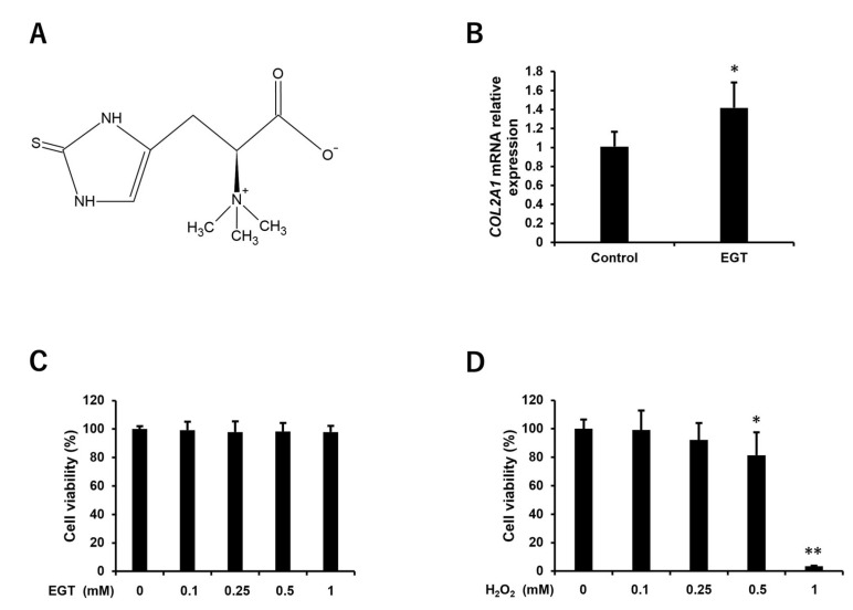
Reactive oxygen species (ROS) induce oxidative stress in cells and are associated with various diseases, including autoimmune diseases. Ergothioneine (EGT) is a natural amino acid derivative derived from the ergot fungus and has been reported to exhibit an effective antioxidant function in many models of oxidative stress-related diseases. Recently, mutations in OCTN1, a membrane transporter of EGT, have been reported to be associated with rheumatoid arthritis. Therefore, we investigated the chondrocyte-protective function of EGT using a model of oxidative stress-induced injury of chondrocytes by hydrogen peroxide (H2O2). Human chondrocytes were subjected to oxidative stress induced by H2O2 treatment, and cell viability, the activity of lactate dehydrogenase (LDH) released into the medium, dead cell ratio, intracellular ROS production, and mitochondrial morphology were assessed. EGT improved chondrocyte viability and LDH activity in the medium and strongly suppressed the dead cell ratio. EGT also exerted protective effects on intracellular ROS production and mitochondrial morphology. These results provide evidence to support the protective effects of EGT on chondrocytes induced by oxidative stress.

Keywords: chondrocyte protection; ergothioneine; oxidative stress; reactive oxygen species; rheumatoid arthritis.


Conflict of interest statement

The authors declare no conflicts of interest.

Figures

Figure 1
Figure 1
Effect of EGT and H2O2 on cell viability of human chondrocytes. (A) Structural formula of EGT. (B) Relative gene expression of COL2A1 in chondrocytes treated with EGT (1 mM) for 24 h. COL2A1 mRNA levels were normalized to ACTB mRNA levels. The values are presented as mean ± SD (n = 4). (C) Cell viability of chondrocytes treated with EGT at various concentrations (0–1 mM) for 24 h. (D) Cell viability of chondrocytes treated with H2O2 at various concentrations (0–1 mM) for 24 h. Cell viability was determined by CCK-8 assay. The values of cell viability are presented as mean ± SD (n = 8). * p < 0.05, ** p < 0.01 vs. control group. EGT, ergothioneine; H2O2, hydrogen peroxide; COL2A1, collagen type 2 alpha 1; ACTB, actin beta; CCK-8, Cell Counting Kit-8; SD, standard deviation.
Figure 2
Figure 2
EGT ameliorates H2O2-induced cell damage in chondrocytes. (A) Morphology of chondrocytes treated with H2O2 (1 mM) and EGT (0–1 mM) for 24 h (scale bar: 100 µm). (B) Cell viability quantified by CCK-8 assay. Values are presented as mean ± SD (n = 6). (C) LDH activity of supernatant of chondrocytes treated with H2O2 (1 mM) and EGT (0–1 mM) for 24 h. LDH activity was measured using Cytotoxicity LDH Assay Kit-WST. Values are presented as mean ± SD (n = 6). ** p < 0.01 vs. control group; ## p < 0.01 vs. H2O2-treated group. EGT, ergothioneine; H2O2, hydrogen peroxide; CCK-8, Cell Counting Kit-8; LDH, lactate dehydrogenase; SD, standard deviation.
Figure 3
Figure 3
EGT inhibits H2O2-induced cell death in chondrocytes. (A) Fluorescence image of chondrocytes treated with H2O2 (1 mM) and EGT (1 mM) for 3, 6, and 12 h (scale bar: 100 µm). Dead cells (green) and total cells (red) were double-stained with Diyo-1 and SYTO59 nuclear staining dyes. (B) Quantitative data on the ratio of dead cells to total cells calculated by double-staining. Values are presented as mean ± SD (n = 3). ** p < 0.01 vs. control group; ## p < 0.01 vs. H2O2-treated group. EGT, ergothioneine; H2O2, hydrogen peroxide; SD, standard deviation.
Figure 4
Figure 4
EGT inhibits H2O2-induced intracellular ROS production in chondrocytes. (A) Fluorescence images of chondrocytes treated with H2O2 (1 mM) and EGT (1 mM) for 3, 6, and 12 h in the presence of DCFH-DA dye (scale bar: 100 µm). (B) Quantitative data on the average fluorescence intensity of chondrocytes obtained from fluorescence images. Values are presented as mean ± SD (n = 3). (C) Changes in cell count over time. The values are expressed as 100% of the cell count in each group at 3 h. * p < 0.05, ** p < 0.01 vs. control group; ## p < 0.01 vs. H2O2-treated group. EGT, ergothioneine; H2O2, hydrogen peroxide; ROS, reactive oxygen species; SD, standard deviation; DCFH-DA, dichloro-dihydro-fluorescein diacetate.
Figure 5
Figure 5
EGT inhibits H2O2-induced mitochondrial condensation in chondrocytes. (A) Fluorescence images of chondrocytes treated with H2O2 (1 mM) and EGT (1 mM) for 2 h in the presence of MitoView Green dye (scale bar: 100 µm). (B) Quantitative data on the average fluorescence intensity of chondrocytes obtained from fluorescence images. Values are presented as mean ± SD (n = 3). ** p < 0.01 vs. control group; ## p < 0.01 vs. H2O2-treated group. EGT, ergothioneine; H2O2, hydrogen peroxide; SD, standard deviation.

References

    1. Sies H., Jones D.P. Reactive oxygen species (ROS) as pleiotropic physiological signalling agents. Nat. Rev. Mol. Cell Biol. 2020;21:363–383. doi: 10.1038/s41580-020-0230-3. - DOI - PubMed
    1. Forman H.J., Zhang H. Targeting oxidative stress in disease: Promise and limitations of antioxidant therapy. Nat. Rev. Drug Discov. 2021;20:689–709. doi: 10.1038/s41573-021-00233-1. - DOI - PMC - PubMed
    1. Hayes J.D., Dinkova-Kostova A.T., Tew K.D. Oxidative stress in cancer. Cancer Cell. 2020;38:167–197. doi: 10.1016/j.ccell.2020.06.001. - DOI - PMC - PubMed
    1. Touyz R.M., Rios F.J., Alves-Lopes R., Neves K.B., Camargo L.L., Montezano A.C. Oxidative stress: A unifying paradigm in hypertension. Can. J. Cardiol. 2020;36:659–670. doi: 10.1016/j.cjca.2020.02.081. - DOI - PMC - PubMed
    1. Yang X., Li Y., Li Y., Ren X., Zhang X., Hu D., Gao Y., Xing Y., Shang H. Oxidative stress-mediated atherosclerosis: Mechanisms and therapies. Front. Physiol. 2017;8:600. doi: 10.3389/fphys.2017.00600. - DOI - PMC - PubMed

Grants and funding

LinkOut - more resources