. 2024 Jul 21;13(7):875.
doi: 10.3390/antiox13070875.

Rod-Shaped Mesoporous Zinc-Containing Bioactive Glass Nanoparticles: Structural, Physico-Chemical, Antioxidant, and Immuno-Regulation Properties

Affiliations

Rod-Shaped Mesoporous Zinc-Containing Bioactive Glass Nanoparticles: Structural, Physico-Chemical, Antioxidant, and Immuno-Regulation Properties

Xiuan Zhu et al. Antioxidants (Basel). .

Abstract

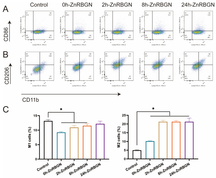
Bioactive glass nanoparticles (BGNs) are applied widely in tissue regeneration. Varied micro/nanostructures and components of BGNs have been designed for different applications. In the present study, nanorod-shaped mesoporous zinc-containing bioactive glass nanoparticles (ZnRBGNs) were designed and developed to form the bioactive content of composite materials for hard/soft tissue repair and regeneration. The nanostructure and components of the ZnRBGNs were characterized, as were their cytocompatibility and radical-scavenging activity in the presence/absence of cells and their ability to modulate macrophage polarization. The ZnRBGNs possessed a uniform rod shape (length ≈ 500 nm; width ≈ 150 nm) with a mesoporous structure (diameter ≈ 2.4 nm). The leaching liquid of the nanorods at a concentration below 0.5 mg/mL resulted in no cytotoxicity. More significant improvements in the antioxidant and M1-polarization-inhibiting effects and the promotion of M2 polarization were found when culturing the cells with the ZnRBGNs compared to when culturing them with the RBGNs. The doping of the Zn element in RBGNs may lead to improved antioxidant and anti-inflammatory effects, which may be beneficial in tissue regeneration/repair.

Keywords: bioactive glass; macrophage polarization; radical-scavenging activity; rod-shaped nanoparticles.


Conflict of interest statement

The authors declare that they have no conflicts of interest.

Figures

Figure 1
Figure 1
Scanning electron microscopy (SEM) images of 0h-ZnRBGN, 2h-ZnRBGN, 8h-ZnRBGN, and 24h-ZnRBGN (×5 k, ×10 k, and ×20 k).
Figure 2
Figure 2
(A) Transmission electron microscope (TEM) images of 0h-ZnRBGN, 2h-ZnBGN, 8h-ZnRBGN, and 24h-ZnRBGN. (B) Energy spectrum analysis and element distribution (Green pots represented distribution of Si element, red for O and blue for Zn) of 0h-ZnRBGN, 2h-ZnRBGN, 8h-ZnRBGN, and 24h-ZnRBGN.
Figure 3
Figure 3
(A) XRD pattern, (B) FTIR, (C) XPS wide scan, and (D) narrow scan analyses of 0h-ZnRBGN, 2h-ZnBGN, 8h-ZnRBGN, and 24h-ZnRBGN.
Figure 4
Figure 4
N2 adsorption–desorption isotherms (A) and pore size distributions (B) of 0h-ZnRBGN, 2h-ZnRBGN, 8h-ZnRBGN, and 24h-ZnRBGN.
Figure 5
Figure 5
The cytotoxicity evaluation for 0h-ZnRBGN, 2h-ZnRBGN, 8h-ZnRBGN, and 24h-ZnRBGN. (A) Live and dead staining. Fluorescence imaging of calcein and PI staining of samples in 0.1 mg/mL. (B) OD value of L929 culture with extract of different samples in 0.01, 0.1, 0.5, and 1 mg/mL. (C) Quantitative evaluation of live and dead staining. * p < 0.05, “ns” means no significant difference.
Figure 6
Figure 6
Evaluation of the effects of 0.1 mg/mL extracts of 0h-ZnRBGN, 2h-ZnRBGN, 8h-ZnRBGN, and 24h-ZnRBGN proliferation. (A) EDU and Dapi staining. The EDU fluorescence imaging and Dapi staining for the 0.1 mg/mL samples. (B) The quantitative evaluation of staining. (C) The OD values of the L929 culture with 0.1 mg/mL extracts of the different samples on days 1 and 3, “ns” means no significant difference.
Figure 7
Figure 7
The flow cytometry analysis of macrophage surface marker expression; the fluorescence intensity of CD86 (A) and CD206 (B) of the RAW 264.7 cells cultured with 0h-ZnRBGN, 2h-ZnRBGN, 8h-ZnRBGN, and 24h-ZnRBGN. (C) The macrophage populations of M1 (CD86)- and M2-polarized (CD206) RAW 264.7 cells with 0h-ZnRBGN, 2h-ZnRBGN, 8h-ZnRBGN, and 24h-ZnRBGN. * p < 0.05.
Figure 8
Figure 8
Antioxidant effect analysis. (A) ROS generation assay analysis. Images of the cells cultured with the leaching medium (green fluorescence represents ROS) and quantitative analysis (B). (C) The DPPH free radical-scavenging rates with 0h-ZnRBGN and 8h-ZnRBGN. * p < 0.05, “ns” means no significant difference.

References

    1. Francesco B., Sepideh H., Saeid K. Bioactive glasses: Where Are We and Where Are We Going. J. Func. Biomater. 2018;9:25. doi: 10.3390/jfb9010025. - DOI
    1. Denissen H.W., Groot D.K., Makkes P.C., Hooff V.D., Klopper P.J. Tissue Response to Dense Apatite Implants in Rats. J. Biomed. Mater. Res. 1980;14:713–721. doi: 10.1002/jbm.820140603. - DOI - PubMed
    1. Vallet-Regi M., Salinas A.J. Mesoporous Bioactive Glasses for Regenerative Medicine. Mater. Today Bio. 2021;11:100121. doi: 10.1016/j.mtbio.2021.100121. - DOI - PMC - PubMed
    1. Charlotte V., Jean-Marie N. Bioactive Glass Nanoparticles: From Synthesis to Materials Design for Biomedical Applications. Materials. 2016;9:288. doi: 10.3390/ma9040288. doi: 10.3390/ma9040288. - DOI - DOI - PMC - PubMed
    1. Kozon D., Zheng K., Boccardi E., Liu Y., Liverani L., Boccaccini A.R. Synthesis of Monodispersed Ag-Doped Bioactive Glass Nanoparticles via Surface Modification. Materials. 2016;9:225. doi: 10.3390/ma9040225. - DOI - PMC - PubMed

LinkOut - more resources