

Spontaneous Reduction in the Intermetatarsal Angle in Distal First Metatarsal Osteotomies with No Lateral Head Displacement in Hallux Valgus
- PMID: 39062011
- PMCID: PMC11275205
- DOI: 10.3390/biomedicines12071438
Spontaneous Reduction in the Intermetatarsal Angle in Distal First Metatarsal Osteotomies with No Lateral Head Displacement in Hallux Valgus
Abstract
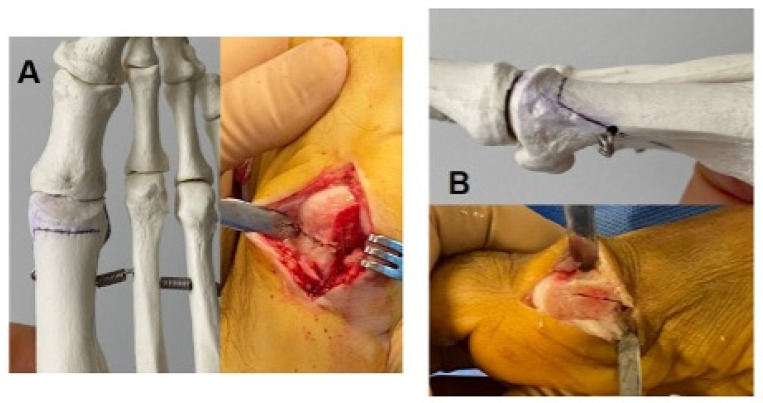
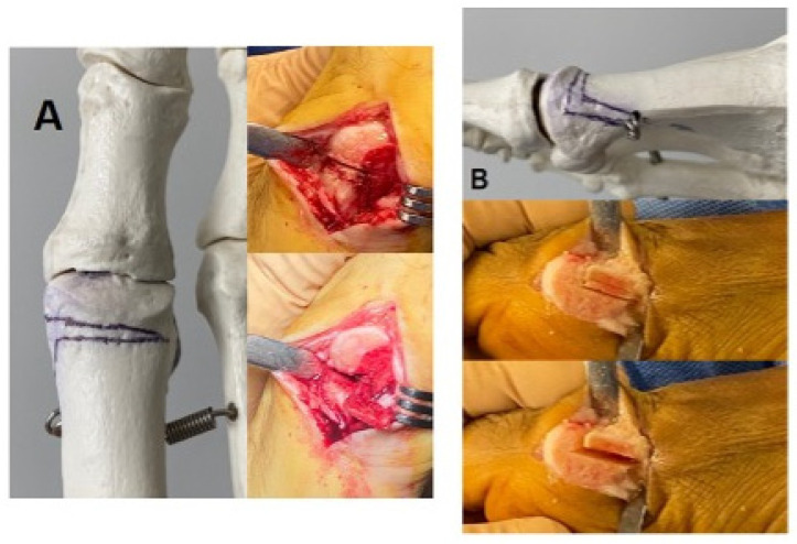
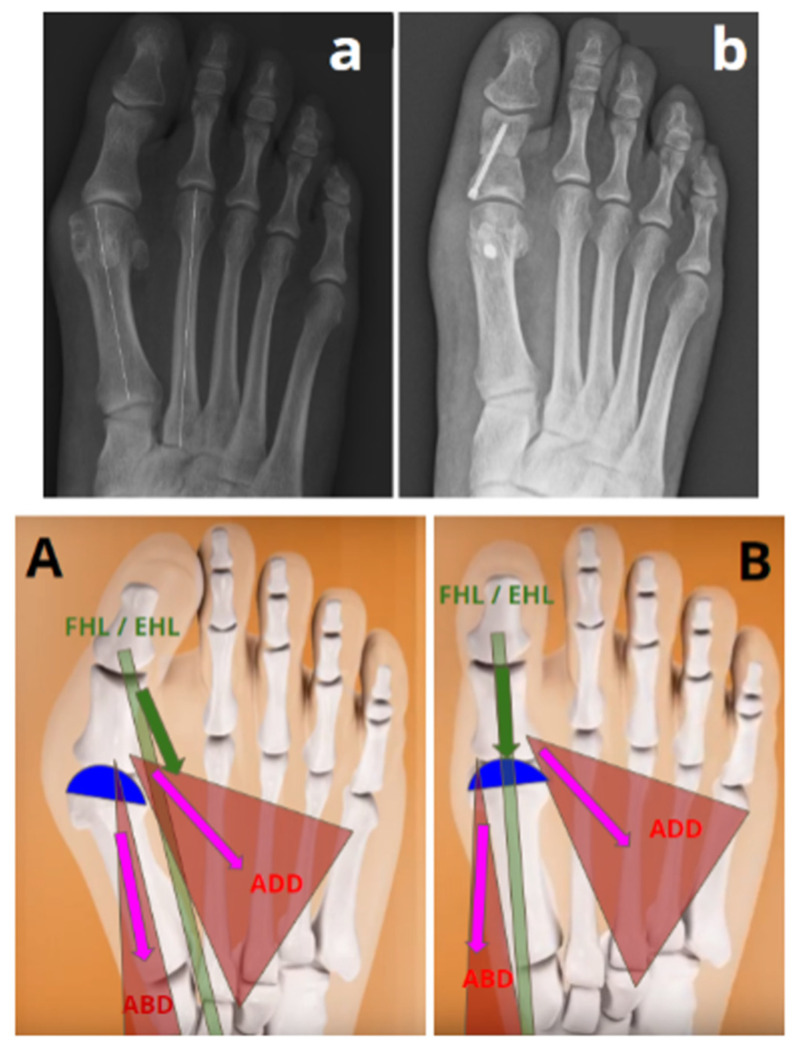
Background: The outcomes of first metatarsal (M1) distal osteotomies in hallux valgus (HV) can be improved, especially for intermetatarsal angle (IMA) correction, which is mainly based on lateral displacement of the M1 head (i.e., translation) through the osteotomy. Conversely, there is a spontaneous reduction in the IMA in first metatarsophalangeal joint (MTP1) arthrodesis. But we do not know whether this can be applied to distal osteotomies. We propose a distal osteotomy, called 3D chevron, which combines supination and varization of the M1 head. This might realign soft tissues around the MTP1, potentially leading to a spontaneous reduction in the IMA by an analogous mechanism to MTP1 fusion. Therefore, our study aimed to assess whether spontaneous reductions in IMAs exist in distal M1 osteotomies in the absence of lateral translations of M1 heads.
Methods: A prospective continuous series of 25 3D chevrons was performed. Two groups were formed during surgery. Patients requiring no M1 head lateral displacement were included in the "successful correction without translation" group, and patients requiring M1 head lateral displacement were included in the "failed correction without translation" group. Radiographic analysis was performed preoperatively and at 1 year postoperatively.
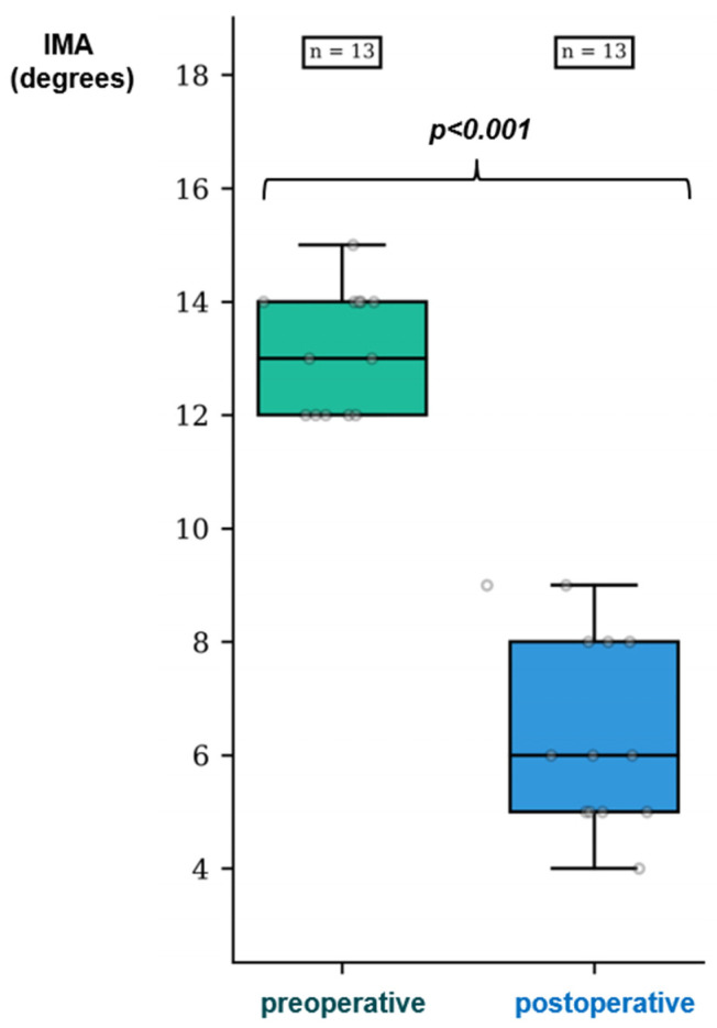
Results: Twenty-two women and three men, with a mean age of 44.8 ± 14.2 years and a mean body mass index of 22.6 ± 4.1 kg/m2, underwent follow-up at one year after surgery. The "successful correction without translation" group was composed of HV with milder deformities (13/25 HVs, median preoperative IMA = 13 (IQR 2)) compared to the "failed correction without translation" group (median IMA = 16 (IQR 2.25) p < 0.001). Spontaneous reductions in IMAs were observed in the "successful correction without translation" group, with a median decrease in the IMA of 6 degrees (CI95%[5.5; 8.0]; p < 0.001) between preoperative and 1-year radiographs.
Conclusion: Distal osteotomies allow for spontaneous reduction in the IMA in HV. First metatarsal head translation through an osteotomy should not be considered as the only procedure to correct IMAs in distal osteotomies.
Keywords: distal metatarsal articular angle; hallux valgus; intermetatarsal angle; pronation; supination.
Conflict of interest statement
The authors declare no conflicts of interest.
Figures
Similar articles
-
[Surgical management of hallux valgus by techniques preserving the first metatarsophalangeal joint: long-term results].Acta Chir Orthop Traumatol Cech. 2007 Apr;74(2):105-10. Acta Chir Orthop Traumatol Cech. 2007. PMID: 17493411 Czech.
-
Double First Metatarsal and Akin Osteotomy for Severe Hallux Valgus.Foot Ankle Int. 2015 Oct;36(10):1215-22. doi: 10.1177/1071100715589173. Epub 2015 Jun 24. Foot Ankle Int. 2015. PMID: 26109608
-
Impact of First Metatarsal Hyperpronation on First Ray Alignment: A Study in Cadavers.Clin Orthop Relat Res. 2022 Oct 1;480(10):2029-2040. doi: 10.1097/CORR.0000000000002265. Epub 2022 Jun 7. Clin Orthop Relat Res. 2022. PMID: 35700368 Free PMC article.
-
Proximal versus distal metatarsal osteotomies for moderate to severe hallux valgus deformity: a systematic review and meta-analysis of clinical and radiological outcomes.Int Orthop. 2018 Aug;42(8):1853-1863. doi: 10.1007/s00264-018-3782-5. Epub 2018 Feb 10. Int Orthop. 2018. PMID: 29427126
-
Comparison of outcomes of different osteotomy sites for hallux valgus: A systematic review and meta-analysis.J Orthop Surg (Hong Kong). 2022 May-Aug;30(2):10225536221110473. doi: 10.1177/10225536221110473. J Orthop Surg (Hong Kong). 2022. PMID: 35836406
Cited by
-
Valgus Deviation of the Intersesamoid Crista in Hallux Valgus and Its Association With the Distal Metatarsal Articular Angle: A Pilot Study.Foot Ankle Orthop. 2025 Aug 16;10(3):24730114251355501. doi: 10.1177/24730114251355501. eCollection 2025 Jul. Foot Ankle Orthop. 2025. PMID: 40827130 Free PMC article.
References
-
- Santos M., Roseiro L., Seiça E.C., Amaro A.M. A Systematic Review of Osteotomies to Correct Hallux Valgus in the First Metatarsal. Appl. Sci. 2024;14:3043. doi: 10.3390/app14073043. - DOI
-
- Lalevee M., de Cesar Netto C., ReSurg, Boublil D., Coillard J.Y. Recurrence Rates with Longer-Term Follow-up After Hallux Valgus Surgical Treatment with Distal Metatarsal Osteotomies: A Systematic Review and Meta-Analysis. Foot Ankle Int. 2023;44:210–222. doi: 10.1177/10711007231152487. - DOI - PubMed
LinkOut - more resources
Full Text Sources
