. 2024 Jun 27;12(7):1439.
doi: 10.3390/biomedicines12071439.

Suppressing Pro-Apoptotic Proteins by siRNA in Corneal Endothelial Cells Protects against Cell Death

Affiliations

Suppressing Pro-Apoptotic Proteins by siRNA in Corneal Endothelial Cells Protects against Cell Death

Susanne Staehlke et al. Biomedicines. .

Abstract

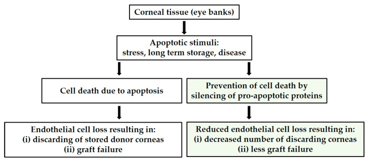
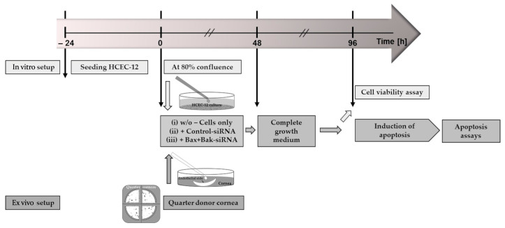
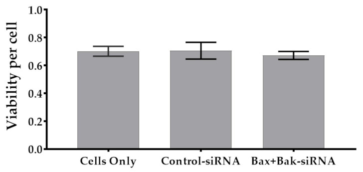
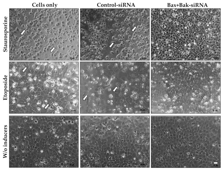
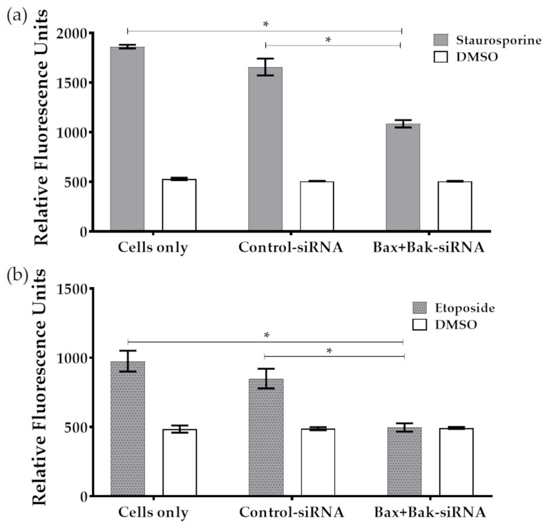
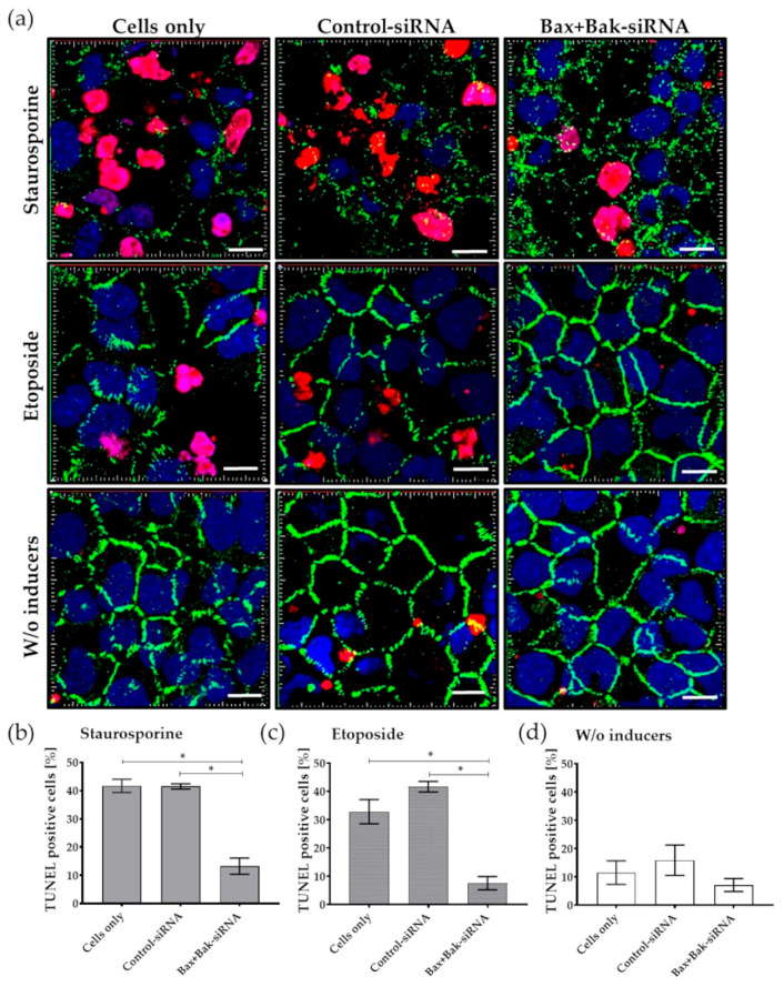
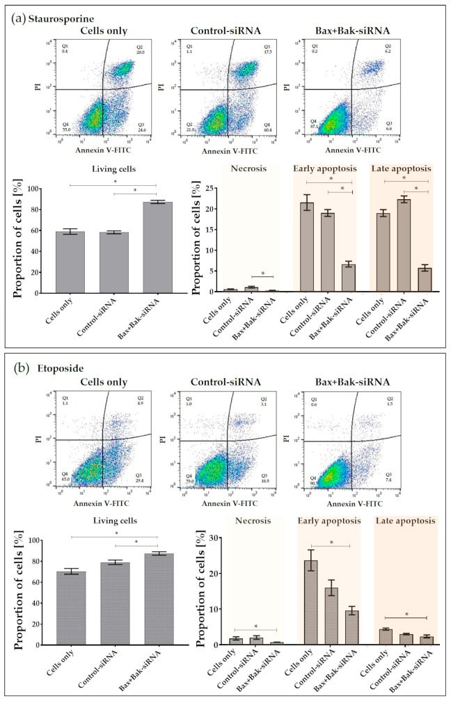
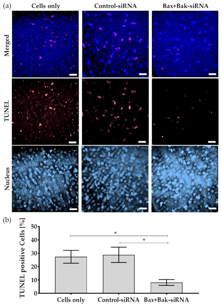
Corneal endothelial cells (CE) are critical for the cornea's transparency. For severe corneal damage, corneal tissue transplantation is the most promising option for restoring vision. However, CE apoptotic cell death occurs during the storage of donor corneas for transplantation. This study used small interfering (si)RNA-mediated silencing of pro-apoptotic proteins as a novel strategy to protect CE against apoptosis. Therefore, the pro-apoptotic proteins Bax and Bak were silenced in the human corneal endothelial cell line (HCEC-12) by transfection with Accell™siRNA without any adverse effects on cell viability. When apoptosis was induced, e.g., etoposide, the caspase-3 activity and Annexin V-FITC/PI assay indicated a significantly reduced apoptosis rate in Bax+Bak-siRNA transfected HCECs compared to control (w/o siRNA). TUNEL assay in HCECs exposed also significantly lower cell death in Bax+Bak-siRNA (7.5%) compared to control (w/o siRNA: 32.8%). In ex vivo donor corneas, a significant reduction of TUNEL-positive CEs in Bax+Bak-siRNA corneas (8.1%) was detectable compared to control-treated corneas (w/o siRNA: 27.9%). In this study, we demonstrated that suppressing pro-apoptotic siRNA leads to inhibiting CE apoptosis. Gene therapy with siRNA may open a new translational approach for corneal tissue treatment in the eye bank before transplantation, leading to graft protection and prolonged graft survival.

Keywords: Annexin V; TUNEL; anti-apoptotic Bax and Bak; apoptosis; caspase-3; confocal laser scanning microscopy; corneal endothelial cells; donor cornea; siRNA.


Conflict of interest statement

The authors declare no conflicts of interest.

Figures

Figure 1
Figure 1
Chart illustrating the causes of endothelial cell loss leading to tissue wastage and the proposed strategy to circumvent this wastage.
Figure 2
Figure 2
Schematic representation of the time course of the experiments. For the in vitro approach, corneal endothelial cells (HCECs) were seeded for 24 h at a density of 2 × 10⁵ to reach 80% confluence for the experiment. For the ex vivo approach, the donor cornea was quartered, and one quarter was placed with the endothelial side up in a well (“cornea in a cup”). The procedure for both cultures (highlighted in green) was then to apply siRNA in serum-free medium at time 0 h with the following treatment: (i) no addition of siRNA as control (Cells only); (ii) addition of non-target siRNA (Control-siRNA); or addition of siRNA to knockdown the pro-apoptotic proteins Bax and Bak (Bax+Bak-siRNA). After 48 h, the medium in both cultures was changed, and the appropriate complete growth medium was added. After a further 48 h of knockdown, experiments were performed for the HCEC viability assay. In order to analyze the transfection, cell death was induced with etoposides (HCECs additional with staurosporine), followed by apoptosis assays such as TUNEL.
Figure 3
Figure 3
Relative cell viability of corneal endothelial cells (HCECs) after 96-hour transfection procedure. Note that the transfection with Accell siRNA showed no effect on cell viability. Cell metabolism (MTS assay) values were related to cell number (crystal violet). (Anthos reader; n = 4, mean ± s.e.m., Friedmann test posthoc Dunn’s multiple comparison test: not significant).
Figure 4
Figure 4
Cell morphology of human corneal endothelial cells (HCECs) under different conditions: cells were transfected with the Accell siRNA (Control-siRNA, Bax+Bak-siRNA) or untransfected (Cells only) for 96 h (w/o inducers, last row), followed by staurosporine (250 nM for 6 h; first row) or by etoposide treatment (42.5 µM for 21 h; middle row). It can be seen that the induction of apoptosis with both staurosporine and etoposide alters the phenotype of HCECs in controls (Cells only and Control-siRNA). Under staurosporine treatment, the cells shrink significantly and can no longer form cell clusters. The loss of cells and contacts can also be observed with etoposide treatment (arrows). In contrast, Bax+Bak-siRNA transfected HCECs and treated with apoptosis inducers have improved morphology, resembling cells without apoptosis stimulus (w/o inducers). (Axiovert 40C, Zeiss; bars 20 µm).
Figure 5
Figure 5
The extent of apoptosis was measured in terms of caspase-3 activity after induction of apoptosis in transfected (Control-siRNA, Bax+Bak-siRNA) and untransfected (Cells only) HCECs: (a) Apoptosis was induced using Staurosporine (250 nM for 6 h) after 96 h transfection. (b) Apoptosis was induced using etoposide (42.5 µM for 21 h) after 96 h transfection. Note that in both cases, the apoptosis rate was significantly reduced due to the knockdown of pro-apoptotic proteins Bax and Bak. (microplate reader Fluoroskan Ascent FL; n = 3, mean ± s.e.m., Friedman-Test posthoc Dunn’s multiple comparison test: * p < 0.05).
Figure 6
Figure 6
Detection of apoptosis rate by TUNEL assay in human corneal endothelial cells (HCECs) after transfection with Accell Control-siRNA or Bax+Bak-siRNA compared to untransfected cells (Cells only). After incubation for 96 h, the apoptosis inducers staurosporine (250 nM for 6 h) or etoposide (42.5 µM for 21 h) were added. (a) Exemplary images of apoptosis rate and changes in cell morphology in HCECs labeled with TUNEL assay and staining of cell-cell contacts (ZO-1). Note that due to the induction of apoptosis, many nuclei of apoptotic cells (red) and gaps in the cell-cell barrier (green) could be detected in controls—Cells only and Control-siRNA. In contrast, almost no TUNEL-positive cells are found in Bax+Bak-siRNA-transfected cells, and tight cell-cell contact of HCECs can be observed, comparable to endothelial cells without (w/o) apoptosis induction for etoposide and to a lesser extent for staurosporine. (LSM780, Zeiss; red: TUNEL—apoptotic DNA fragmentation, blue: Dapi—cell nuclei, green: ZO-1—cell-cell contacts; 63× oil objective, zoom 2, 3D overlay, bar = 10 µm). Images were taken from five random fields, TUNEL-positive cells were counted, and the percentage for (b) staurosporine, (c) etoposide, and (d) cells treated without inducers was calculated. Quantification clearly shows that the apoptosis rate was reduced after the knockdown of Bax+Bak. (ImageJ; n = 3, mean ± s.e.m., RM one-way ANOVA posthoc Bonferroni’s multiple comparison test: * p < 0.05).
Figure 7
Figure 7
Detection of apoptosis rate using Annexin V-FITC/Propidium Iodide (PI) in human corneal endothelial cells (HCECs) after transfection with Accell Control-siRNA or Bax+Bak-siRNA compared to untransfected cells (Cells only). After incubation for 96 h, cells were incubated with either apoptosis inducers (a) staurosporine (250 nM for 6 h) or (b) etoposide (42.5 µM for 21 h). Representative flow cytometric analysis of HCECs under the respective conditions. (FACSCalibur, BD; cells in Q1: necrosis, Q2: late apoptosis, Q3: early apoptosis; Q4: live; FL-1: Annexin, FL-2: PI). Quantification shows that significantly more live cells were present after the Bax+Bak knockdown. (FlowJo; n = 3, mean ± s.e.m., RM one-way ANOVA posthoc uncorrected Fisher’s LSD test: * p < 0.05).
Figure 8
Figure 8
As a proof-of-principle, apoptosis was detected by TUNEL (TdT-mediated dUTP-biotin nick end labeling) assay in the endothelium of donor corneas after transfection with Accell Control-siRNA or Bax+Bak-siRNA compared to untransfected cells (Cells only). After incubation for 96 h, the apoptosis inducer etoposide (42.5 µM for 21 h) was added. (a) Exemplary images of the nuclei of the cells labeled with TUNEL (red) and Dapi (blue). Note that despite the induction of apoptosis, fewer nuclei of apoptotic cells were visible in the corneal endothelium compared to controls—Cells only and Control-siRNA. (LSM780, Zeiss; red: TUNEL—apoptotic DNA fragmentation; blue: Dapi—cell nuclei; 40× objective, zoom 0.6, 3D overlay, bar = 40 µm). (b) Quantification indicated that the apoptosis rate was significantly reduced in corneas after the Bax+Bak knockdown. (ImageJ; n = 3, mean ± s.e.m., RM one-way ANOVA posthoc uncorrected Fisher’s LSD test: * p < 0.05).

Similar articles

References

    1. Kreutziger G.O. Lateral membrane morphology and gap junction structure in rabbit corneal endothelium. Exp. Eye Res. 1976;23:285–293. doi: 10.1016/0014-4835(76)90129-9. - DOI - PubMed
    1. Waring G.O., 3rd, Bourne W.M., Edelhauser H.F., Kenyon K.R. The corneal endothelium. Normal and pathologic structure and function. Ophthalmology. 1982;89:531–590. doi: 10.1016/S0161-6420(82)34746-6. - DOI - PubMed
    1. Capella J.A. Regeneration of endothelium in diseased and injured cornea. Am. J. Ophthalmol. 1972;74:810–817. doi: 10.1016/0002-9394(72)91200-7. - DOI - PubMed
    1. Zhu C., Joyce N.C. Proliferative response of corneal endothelial cells from young and older donors. Investig. Ophthalmol. Vis. Sci. 2004;45:1743–1751. doi: 10.1167/iovs.03-0814. - DOI - PubMed
    1. Altamirano F., Ortiz-Morales G., O’Connor-Cordova M.A., Sancén-Herrera J.P., Zavala J., Valdez-Garcia J.E. Fuchs endothelial corneal dystrophy: An updated review. Int. Ophthalmol. 2024;44:61. doi: 10.1007/s10792-024-02994-1. - DOI - PubMed

LinkOut - more resources