Review
. 2024 Jul 9;12(7):1527.
doi: 10.3390/biomedicines12071527.

Osteopontin: A Key Multifaceted Regulator in Tumor Progression and Immunomodulation

Affiliations
Review

Osteopontin: A Key Multifaceted Regulator in Tumor Progression and Immunomodulation

Venketesh K Panda et al. Biomedicines. .

Abstract

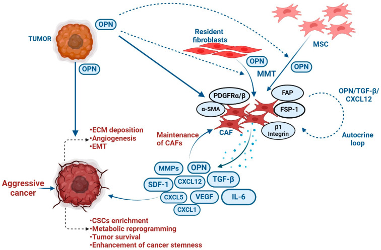
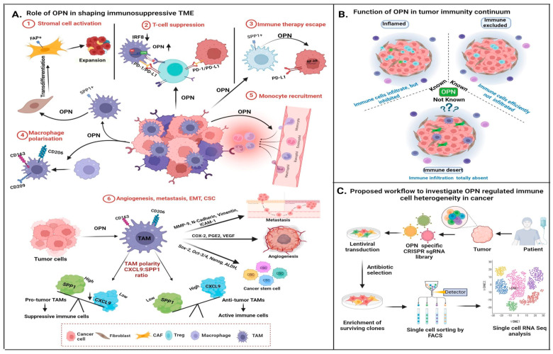
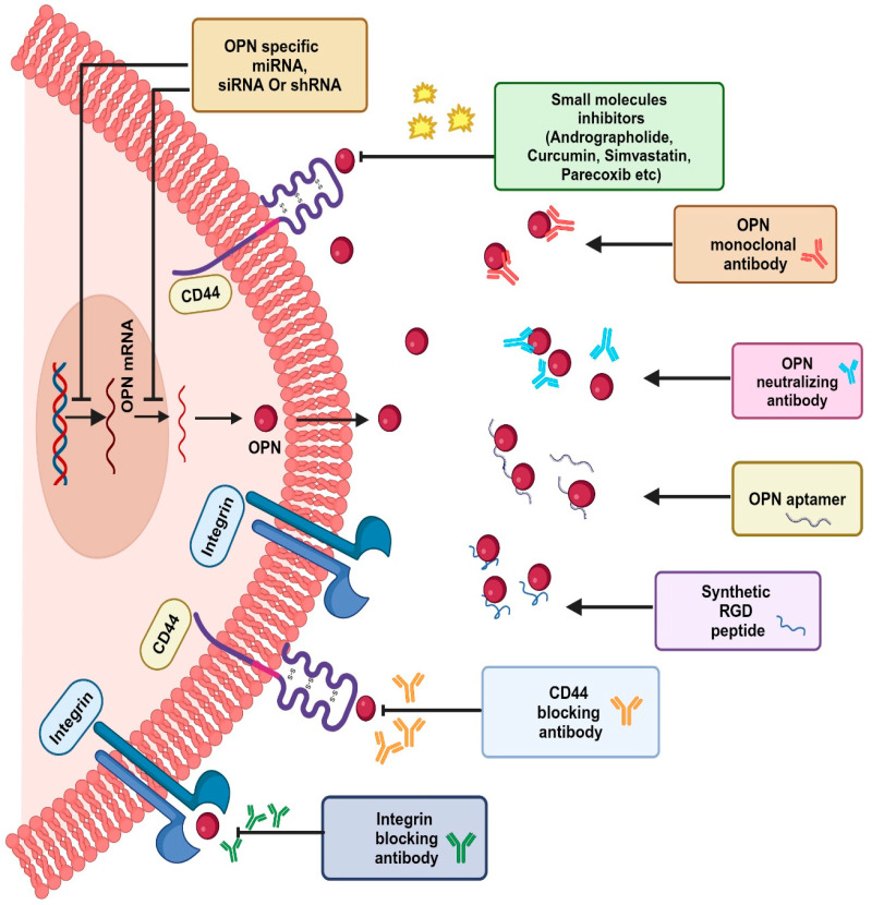
The tumor microenvironment (TME) is composed of various cellular components such as tumor cells, stromal cells including fibroblasts, adipocytes, mast cells, lymphatic vascular cells and infiltrating immune cells, macrophages, dendritic cells and lymphocytes. The intricate interplay between these cells influences tumor growth, metastasis and therapy failure. Significant advancements in breast cancer therapy have resulted in a substantial decrease in mortality. However, existing cancer treatments frequently result in toxicity and nonspecific side effects. Therefore, improving targeted drug delivery and increasing the efficacy of drugs is crucial for enhancing treatment outcome and reducing the burden of toxicity. In this review, we have provided an overview of how tumor and stroma-derived osteopontin (OPN) plays a key role in regulating the oncogenic potential of various cancers including breast. Next, we dissected the signaling network by which OPN regulates tumor progression through interaction with selective integrins and CD44 receptors. This review addresses the latest advancements in the roles of splice variants of OPN in cancer progression and OPN-mediated tumor-stromal interaction, EMT, CSC enhancement, immunomodulation, metastasis, chemoresistance and metabolic reprogramming, and further suggests that OPN might be a potential therapeutic target and prognostic biomarker for the evolving landscape of cancer management.

Keywords: cancer; cancer-associated fibroblasts; immunomodulation; osteopontin (OPN); single cell transcriptomics; targeted therapy; tumor-associated macrophages.


Conflict of interest statement

The authors declare that they have no conflict of interest.

Figures

Figure 1
Figure 1
Structural domains of full-length OPN and its receptors. N-terminus of OPN consists of the poly-D region, calcium-binding domain and ELVTDFPTDLPAT sequence motif, which interacts with α4β1 integrin. The central region consists of the RGD domain, which binds with other integrins such as αvβ3, αvβ1, αvβ5, αvβ6, αvβ8, α5β1 and α8β1; the SVVYGLR sequence binds to α9β1, α4β1 and α4β7 integrins. The C-terminal region includes another calcium-binding domain, MMP-cleavage site and heparin-binding domain, which facilitate the interaction of OPN with CD44.
Figure 2
Figure 2
Schematic illustration of the OPN gene and its splice variants. Alternative splicing of OPN transcript results in five splice variants, which are denoted as OPN-a, OPN-b, OPN-c, OPN-4 and OPN-5. OPN-a is a full-length variant that consists of 7 exons; OPN-b lacks exon 5, while in OPN-c exon 4 is absent. In OPN-4, both exon 4 and exon 5 are missing, whereas OPN-5 is the longest variant, which consists of an additional exon, generated from a portion of intron 3. Additionally, four new sub-variants of OPN-5 (OPN-5b, OPN-5c, OPN-5d, OPN-5e) have been identified. OPN-5a is the same as OPN-5; OPN-5b has the extra shortened exon while OPN-5c has an additional 9 base pairs in the 3’ region of the extra exon. In OPN-5d, there is a deletion of exon 5 with the addition of 9 base pairs in the 3’ region of the extra exon. OPN-5e lacks exon 5.
Figure 3
Figure 3
Role of OPN in the regulation of various signaling pathways. OPN through its interaction with αvβ3, α4β1, α3β2 and α9β1 integrins and the CD44 receptor transduces multiple signaling pathways and their crosstalks such as FAK/MEK/ERK, PLCγ/PKC/PI3K/Akt/mTOR, NIK/IκBα/NFκB, JAK/STAT3, PI3K/Akt/β-catenin, NFκB/HIF1α/BMI1, c-Src/EGFR/MEK/ERK and MAPK pathways. These signaling cascades induce the activation of various tumor-promoting genes such as VEGF, MMPs and COX-2, thereby inducing tumor growth at the primary sites, angiogenesis, metastases at the distance sites, ECM remodeling, immune suppression, stemness, immune evasion, chemoresistance, migration and survival.
Figure 4
Figure 4
Diagrammatic representation of multifaceted function of OPN in various tumors. OPN regulates EMT, resulting in the loss of tight junctions, thereby enhancing metastasis with the low expression of E-cadherin and high expressions of N-cadherin, vimentin, slug, snail and fibronectin. Under hypoxic conditions, OPN induces activation of PI3K leading to phosphorylation of Akt, thereby upregulating VEGF-dependent angiogenesis. OPN is responsible for metabolic function by activating HIF1α under hypoxic conditions, which further aids in the glycolytic process with high expression of VEGF, PDK1, LDHA, iNOS, GLUT1 and GLUT3. OPN further regulates CSC enrichment by activating a cascade of signaling pathways involving PI3K/Akt/mTOR, hedgehog, MAPK, Wnt/β-catenin, JAK/STAT and notch signaling. The interaction between OPN, αvβ3 and CD44 results in the activation of PI3K/Akt, FAK/MEK/ERK, EGFR and Wnt/NFκB signaling cascades, thereby aiding therapeutic resistance.
Figure 5
Figure 5
Differentiation of resident fibroblasts and MSCs into myofibroblasts by tumor-derived OPN. Tumor-derived OPN is involved in the transition of resident fibroblasts and MSCs into myofibroblast or CAFs. CAF-derived factors induce ECM deposition, EMT, angiogenesis, CSC enrichment, metabolic reprogramming and tumor survival, resulting in the enhancement of tumor progression.
Figure 6
Figure 6
Model depicting the role of OPN in tumor immune microenvironment (TIME): (A) Role of OPN in shaping immunosuppressive TME: tumor-derived OPN activates stromal cells by trans-differentiation of fibroblasts to myofibroblasts, resulting in expansion of tumor. OPN-regulated PD-1/PDL1 interaction inhibits T-cell activation. Further, tumor cells downregulate the expression of IRF8, resulting in overexpression of OPN, thus leading to T-cell suppression. OPN, via the NF-κB pathway, upregulates PD-L1 expression, aiding in immune therapy escape. OPN induces the polarization of macrophages and the recruitment of monocytes. It activates TAM, leading to angiogenesis, metastasis and enrichment of CSCs via upregulation of various tumor-promoting factors such as MMP-9, N-cadherin, vimentin, ICAM-1, COX-2, PGE-2, VEGF, Sox-2, Oct-3/4, Nanog and ALDH. The polarity ratio of CXCL9 and OPN (SPP1) determines the anti- and pro-tumorigenic properties of TAMs. (B) Involvement of OPN in tumor immunity continuum: OPN is primarily associated with inflamed and immune-excluded tumors, whereas its role in immune-desert tumor remains elusive. (C) Schematic representations to identify OPN-regulated immune cell heterogeneity in cancer: OPN-regulated immune modulatory genes may be identified in TIME by CRISPR technology in breast cancer using scRNA-seq based platform.
Figure 7
Figure 7
OPN-targeted novel therapeutic strategies. OPN-specific siRNA, miRNA, shRNA, Small molecule inhibitors (Andrographolide, Curcumin, etc.), OPN neutralizing antibodies, OPN aptamers, synthetic RGD peptides, and CD44 and integrin blocking antibodies have been used recently as therapeutic approaches to target the OPN-integrin/CD44 axis, which leads to downregulation of various oncogenic molecules and suppression of tumor progression by disrupting the OPN-regulated signaling pathways in cancers.

References

    1. Bray F., Laversanne M., Sung H., Ferlay J., Siegel R.L., Soerjomataram I., Jemal A. Global cancer statistics 2022: GLOBOCAN estimates of incidence and mortality worldwide for 36 cancers in 185 countries. CA Cancer J. Clin. 2024;74:229–263. doi: 10.3322/caac.21834. - DOI - PubMed
    1. Mao X., Xu J., Wang W., Liang C., Hua J., Liu J., Zhang B., Meng Q., Yu X., Shi S. Crosstalk between cancer-associated fibroblasts and immune cells in the tumor microenvironment: New findings and future perspectives. Mol. Cancer. 2021;20:131. doi: 10.1186/s12943-021-01428-1. - DOI - PMC - PubMed
    1. Rangaswami H., Bulbule A., Kundu G.C. Osteopontin: Role in cell signaling and cancer progression. Trends Cell Biol. 2006;16:79–87. doi: 10.1016/j.tcb.2005.12.005. - DOI - PubMed
    1. Cho H.-J., Cho H.-J., Kim H.-S. Osteopontin: A multifunctional protein at the crossroads of inflammation, atherosclerosis, and vascular calcification. Curr. Atheroscler. Rep. 2009;11:206–213. doi: 10.1007/s11883-009-0032-8. - DOI - PubMed
    1. Zhao H., Chen Q., Alam A., Cui J., Suen K.C., Soo A.P., Eguchi S., Gu J., Ma D. The role of osteopontin in the progression of solid organ tumour. Cell Death Dis. 2018;9:356. doi: 10.1038/s41419-018-0391-6. - DOI - PMC - PubMed

LinkOut - more resources