. 2024 Jul 16;12(7):1577.
doi: 10.3390/biomedicines12071577.

Simultaneous Expression of Different Therapeutic Genes by Infection with Multiple Oncolytic HSV-1 Vectors

Affiliations

Simultaneous Expression of Different Therapeutic Genes by Infection with Multiple Oncolytic HSV-1 Vectors

Adriana Vitiello et al. Biomedicines. .

Abstract

Oncolytic viruses (OVs) are anti-cancer therapeutics combining the selective killing of cancer cells with the triggering of an anti-tumoral immune response. The latter effect can be improved by arming OVs with immunomodulatory factors. Due to the heterogeneity of cancer and the tumor microenvironment, it is anticipated that strategies based on the co-expression of multiple therapeutic molecules that interfere with different features of the target malignancy will be more effective than mono-therapies. Here, we show that (i) the simultaneous expression of different proteins in triple-negative breast cancer (TNBC) cells can be achieved through their infection with a combination of OVs based on herpes simplex virus type 1 (oHSV1), each encoding a single transgene. (ii) The level of expressed proteins is dependent on the number of infectious viral particles utilized to challenge tumor cells. (iii) All recombinant viruses exhibited comparable efficacy in the killing of TNBC cells in single and multiple infections and showed similar kinetics of replication. Overall, our results suggest that a strategy based on co-infection with a panel of oHSV1s may represent a promising combinatorial therapeutic approach for TNBC, as well as for other types of solid tumors, that merits further investigation in more advanced in vitro and in vivo models.

Keywords: HSV-1; combinatorial approach; immunotherapy; virotherapy.


Conflict of interest statement

The authors declare no conflicts of interest.

Figures

Figure 1
Figure 1
Δγ34.5 HSV-1 replicates in and kills TNBC cells less efficiently than wild-type HSV-1. MDA-MB-231 cells were infected with wild-type (17+) and γ34.5-deleted (Δγ34.5) HSV-1 at an MOI of 0.01 PFU/cell. (a) After 6 and 72 hpi, infected cells were subjected to indirect immunofluorescence staining for ICP4 by confocal microscopy. Nuclei were stained with DRAQ5. Representative images are reported. Scale bars: 0.1 mm. (b) Cell supernatants were collected at the indicated times and infectious viral particles were determined by titration on Vero cells. The graph shows the mean viral titer in PFU/mL and standard error (SE) from at least three independent experiments. (c) Cell viability was determined by trypan blue staining at 72 hpi. Bars display the mean value of the percentage of live cells from infections compared to uninfected cells from three independent experiments. Error bars represent SE. A Student t test was used for statistical analysis (* p value < 0.05; *** p < 0.001; **** p < 0.0001).
Figure 2
Figure 2
ΔΔ-Fluc replicates in MDA-MB-231 cells and kills them more efficiently than Δγ34.5 HSV-1. (a) Schematic of the bacmid of recombinant virus ΔΔ-Fluc. This construct is deleted of the two ɣ34.5 loci as well as of the Us12 gene and expresses Fluc under the control of the human cytomegalovirus immediate early (IE) promoter (PCMV). The PCMV/Fluc cassette is inserted into the UL55 and UL56 intergenic region of HSV-1. The red “X” indicates the Us12 deletion. (b) MDA-MB-231 cells were infected at an MOI of 0.1 PFU/cell. The graph displays the amounts of infectious virions released in the cell supernatants at indicated times post infection. Bars show the mean values and error bars from titrations performed in triplicate. (c) Cell viability was determined by trypan blue staining at 72 hpi. The graph shows the mean values of the percentage of live cells from infections compared to the uninfected cells from three independent experiments. Error bars represent the SE. A Student t test was used for statistical analysis (* p value < 0.05; *** p < 0.001).
Figure 3
Figure 3
Expression of different transgenes from recombinant viruses in MDA-MB-231 cells. (a) Schematic of the bacmids expressing Fluc, EGFP, and IL12. IL12 S1 and IL12-S2 stand for IL12 subunit 1 (p35) and IL12 subunit 2 (p40), respectively. The red “X” indicates the Us12 deletion (b) MDA-MD-231 cells were infected with the indicated recombinant viruses (MOI = 0.1 PFU/cell). At 72 hpi, cells were stained with an ICP4-specific antibody. After incubation with DRAQ5 to mark nuclei, cells were observed by confocal microscopy. Representative images are shown. Scale bars: 0.1 mm. (c) Cells were harvested at 72 hpi and cell viability was evaluated by trypan blue staining. The graphs display the mean values of the percentage of dead cells compared to uninfected cells from at least three independent experiments. Error bars represent the SE. A Student t test was used for statistical analysis (N.S.: not statistically significant; p > 0.05). The expression of Fluc (d) and EGFP (e) was measured using a multi-label plate reader, while the release of IL12 (f) in tissue culture supernatants was measured by ELISA. Graphs in d-e show data from at least three independent experiments.
Figure 4
Figure 4
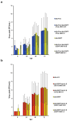
MDA-MB-231 cells infected with a combination of ΔΔ-EGFP and ΔΔ-Fluc express both transgenes and show clear virus-induced cytopathic effects. MDA-MB-231 cells were co-infected with ΔΔ-EGFP and ΔΔ-Fluc combined at an MOI of 0.1 or 0.05 PFU/cell each. As a control, cells were also infected with single recombinant viruses (MOI 0.1 PFU/cell). (a) At the indicated time post infection, cells were examined with a Leica epifluorescence DC100 microscope. Representative images are shown. Scale bars: 0.1 mm. (b) At 72 hpi, cells were harvested and cell viability was determined by trypan blue staining. The graphs display the mean values of the percentage of live cells compared to uninfected cells from three independent experiments. Error bars represent the SE. At the same time, the intensity of Fluc (c) and EGFP (d) signals was measured from infected cells. Graphs in c and d show data from three independent experiments.
Figure 5
Figure 5
MDA-MB-231 cells infected with a combination of ΔΔ-EGFP HSV-1 and ΔΔ-IL12 express both transgenes and show clear virus-induced cytopathic effects. MDA-MB-231 cells were co-infected with ΔΔ-EGFP and ΔΔ-IL12 combined at an MOI of 0.1 or 0.05 PFU/cell each. As a control, cells were also infected with single recombinant viruses (MOI 0.1 PFU/cell). (a) At the indicated times post infection, cells were observed by fluorescence microscopy. Scale bars: 0.1 mm. Representative images are shown. (b) At 72 hpi, cells were harvested and cell viability determined by trypan blue staining. The mean values of the percentage of live cells as compared to uninfected cells from three independent experiments are shown. Error bars represent the SE. At the same time pi, the intensity of the EGFP signal (c) and the amounts of IL12 (pg/mL) released in supernatants of infected MDA-MB-231 (d) were measured. Graphs in c and d show data from three independent experiments.
Figure 6
Figure 6
Recombinant viruses used in combination replicate in MDA-MB-231 as efficiently as in a single infection. MDA-MB 231 cells were co-infected with either ΔΔ-EGFP and ΔΔ-Fluc (a) or with ΔΔ-EGFP and ΔΔ-hIL12 (b) at the indicated MOIs. Single viruses were also adopted for infection as a control at an MOI of 0.1 PFU/cell. At the indicated hpi, cell supernatants were harvested and a plaque assay was performed in Vero cells to evaluate viral titers expressed in PFU/mL. The graphs show the mean and standard errors (SEs) from three independent experiments. Y axis is a logarithmic scale.

References

    1. Tian Y., Xie D., Yang L. Engineering strategies to enhance oncolytic viruses in cancer immunotherapy. Signal Transduct. Target. Ther. 2022;7:117. doi: 10.1038/s41392-022-00951-x. - DOI - PMC - PubMed
    1. Russell S.J., Peng K.W., Bell J.C. Oncolytic virotherapy. Nat. Biotechnol. 2012;30:658–670. doi: 10.1038/nbt.2287. - DOI - PMC - PubMed
    1. Chiocca E.A., Rabkin S.D. Oncolytic viruses and their application to cancer immunotherapy. Cancer Immunol. Res. 2014;2:295–300. doi: 10.1158/2326-6066.CIR-14-0015. - DOI - PMC - PubMed
    1. Kiaheyrati N., Babaei A., Ranji R., Bahadoran E., Taheri S., Farokhpour Z. Cancer therapy with the viral and bacterial pathogens: The past enemies can be considered the present allies. Life Sci. 2024;349:122734. doi: 10.1016/j.lfs.2024.122734. - DOI - PubMed
    1. Shalhout S.Z., Miller D.M., Emerick K.S., Kaufman H.L. Therapy with oncolytic viruses: Progress and challenges. Nat. Rev. Clin. Oncol. 2023;20:160–177. doi: 10.1038/s41571-022-00719-w. - DOI - PubMed

LinkOut - more resources