

Importance of Cardiovascular Magnetic Resonance Applied to Congenital Heart Diseases in Pediatric Age: A Narrative Review
- PMID: 39062326
- PMCID: PMC11276187
- DOI: 10.3390/children11070878
Importance of Cardiovascular Magnetic Resonance Applied to Congenital Heart Diseases in Pediatric Age: A Narrative Review
Abstract
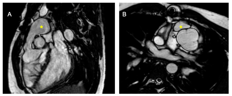
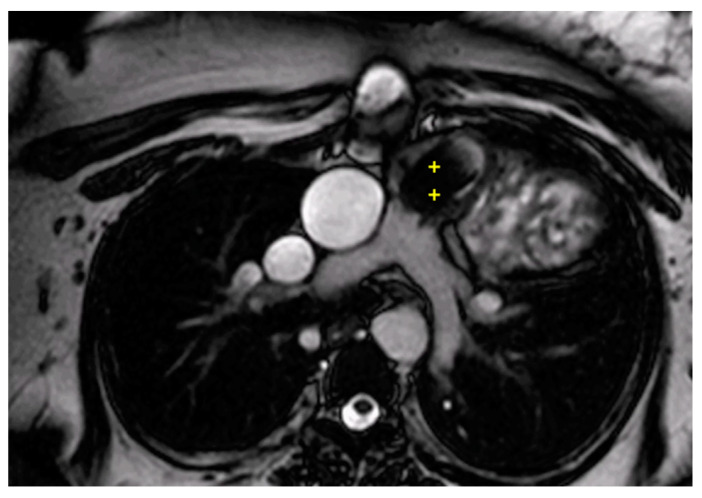
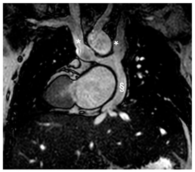
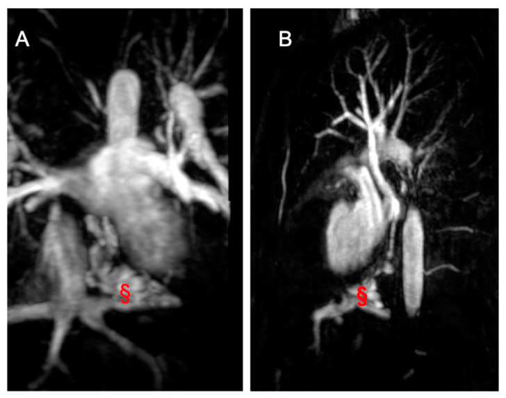
Congenital heart diseases (CHDs) represent a heterogeneous group of congenital defects, with high prevalence worldwide. Non-invasive imaging is essential to guide medical and surgical planning, to follow the patient over time in the evolution of the disease, and to reveal potential complications of the chosen treatment. The application of cardiac magnetic resonance imaging (CMRI) in this population allows for obtaining detailed information on the defects without the necessity of ionizing radiations. This review emphasizes the central role of CMR in the overall assessment of CHDs, considering also the limitations and challenges of this imaging technique. CMR, with the application of two-dimensional (2D) and tri-dimensional (3D) steady-state free precession (SSFP), permits the obtaining of very detailed and accurate images about the cardiac anatomy, global function, and volumes' chambers, giving essential information in the intervention planning and optimal awareness of the postoperative anatomy. Nevertheless, CMR supplies tissue characterization, identifying the presence of fat, fibrosis, or oedema in the myocardial tissue. Using a contrast agent for angiography sequences or 2D/four-dimensional (4D) flows offers information about the vascular, valvular blood flow, and, in general, the cardiovascular system hemodynamics. Furthermore, 3D SSFP CMR acquisitions allow the identification of coronary artery abnormalities as an alternative to invasive angiography and cardiovascular computed tomography (CCT). However, CMR requires expertise in CHDs, and it can be contraindicated in patients with non-conditional devices. Furthermore, its relatively longer acquisition time and the necessity of breath-holding may limit its use, particularly in children under eight years old, sometimes requiring anesthesia. The purpose of this review is to elucidate the application of CMR during the pediatric age.
Keywords: cardiac magnetic technique; congenital heart disease (CHD); magnetic resonance (CMR).
Conflict of interest statement
The authors declare no conflicts of interest.
Figures
Similar articles
-
Cardiac magnetic resonance using fused 3D cine and 4D flow sequences:Validation of ventricular and blood flow measurements.Magn Reson Imaging. 2020 Dec;74:203-212. doi: 10.1016/j.mri.2020.09.026. Epub 2020 Oct 7. Magn Reson Imaging. 2020. PMID: 33035637 Free PMC article.
-
Invasive cardiovascular magnetic resonance (iCMR) for diagnostic right and left heart catheterization using an MR-conditional guidewire and passive visualization in congenital heart disease.J Cardiovasc Magn Reson. 2020 Mar 26;22(1):20. doi: 10.1186/s12968-020-0605-9. J Cardiovasc Magn Reson. 2020. PMID: 32213193 Free PMC article.
-
Multicenter review: role of cardiovascular magnetic resonance in diagnostic evaluation, pre-procedural planning and follow-up for patients with congenital heart disease.Radiol Med. 2016 May;121(5):342-51. doi: 10.1007/s11547-015-0608-z. Epub 2015 Dec 11. Radiol Med. 2016. PMID: 26661952
-
European Association of Cardiovascular Imaging/Cardiovascular Imaging Department of the Brazilian Society of Cardiology recommendations for the use of cardiac imaging to assess and follow patients after heart transplantation.Eur Heart J Cardiovasc Imaging. 2015 Sep;16(9):919-48. doi: 10.1093/ehjci/jev139. Epub 2015 Jul 2. Eur Heart J Cardiovasc Imaging. 2015. PMID: 26139361 Review.
-
Complete Transposition of the Great Arteries in the Pediatric Field: A Multimodality Imaging Approach.Children (Basel). 2024 May 23;11(6):626. doi: 10.3390/children11060626. Children (Basel). 2024. PMID: 38929206 Free PMC article. Review.
Cited by
-
Intracardiac fluid dynamic analysis: available techniques and novel clinical applications.BMC Cardiovasc Disord. 2024 Dec 19;24(1):716. doi: 10.1186/s12872-024-04371-3. BMC Cardiovasc Disord. 2024. PMID: 39702022 Free PMC article. Review.
-
Multimodality Imaging Approach to Infective Endocarditis: Current Opinion in Patients with Congenital Heart Disease.J Clin Med. 2025 Mar 10;14(6):1862. doi: 10.3390/jcm14061862. J Clin Med. 2025. PMID: 40142669 Free PMC article. Review.
References
Publication types
LinkOut - more resources
Full Text Sources
