. 2024 Jun 23;14(7):742.
doi: 10.3390/biom14070742.

Bifidobacterium bifidum SAM-VI Riboswitch Conformation Change Requires Peripheral Helix Formation

Affiliations

Bifidobacterium bifidum SAM-VI Riboswitch Conformation Change Requires Peripheral Helix Formation

Wenwen Xiao et al. Biomolecules. .

Abstract

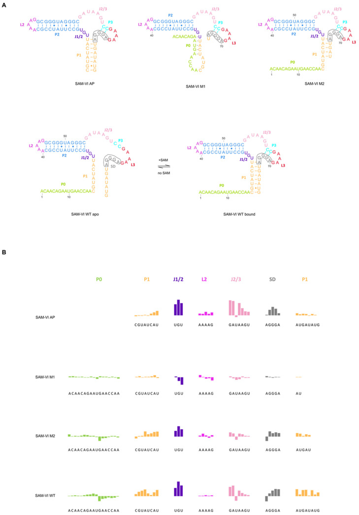
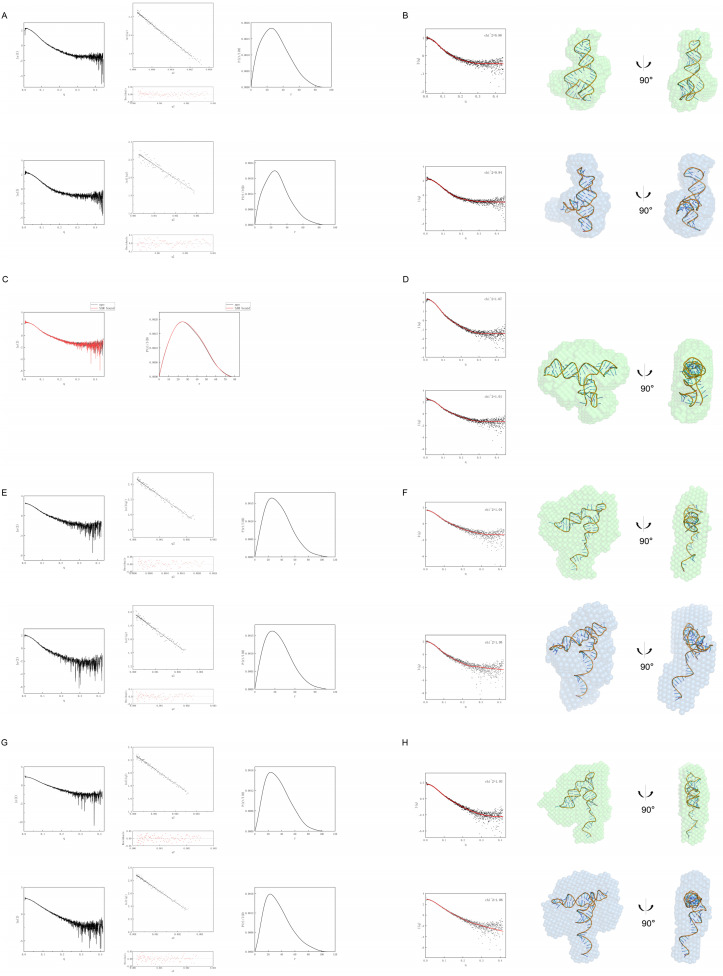
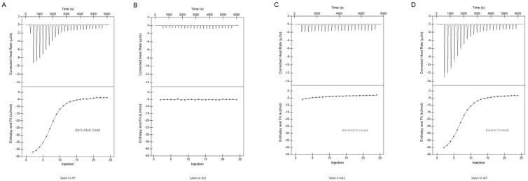
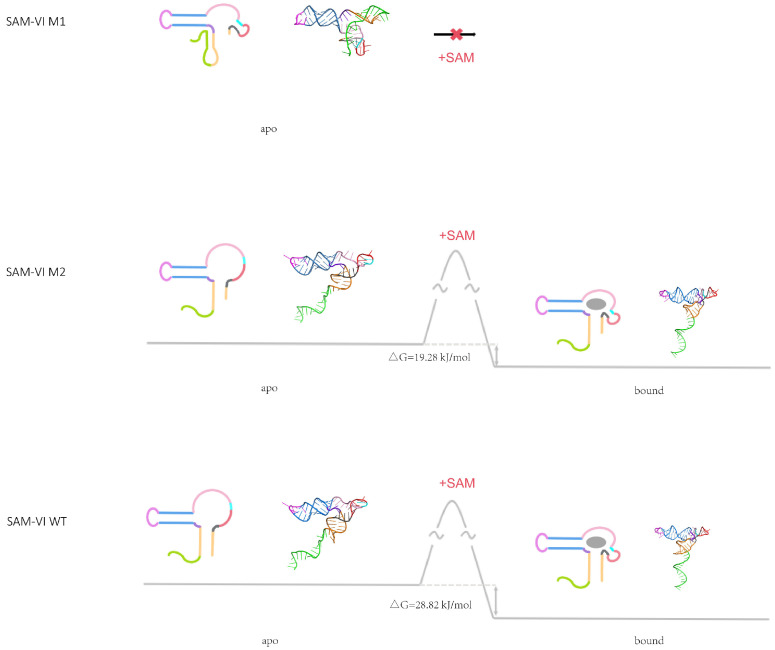
The Bifidobacterium bifidum SAM-VI riboswitch undergoes dynamic conformational changes that modulate downstream gene expression. Traditional structural methods such as crystallography capture the bound conformation at high resolution, and additional efforts would reveal details from the dynamic transition. Here, we revealed a transcription-dependent conformation model for Bifidobacterium bifidum SAM-VI riboswitch. In this study, we combine small-angle X-ray scattering, chemical probing, and isothermal titration calorimetry to unveil the ligand-binding properties and conformational changes of the Bifidobacterium bifidum SAM-VI riboswitch and its variants. Our results suggest that the SAM-VI riboswitch contains a pre-organized ligand-binding pocket and stabilizes into the bound conformation upon binding to SAM. Whether the P1 stem formed and variations in length critically influence the conformational dynamics of the SAM-VI riboswitch. Our study provides the basis for artificially engineering the riboswitch by manipulating its peripheral sequences without modifying the SAM-binding core.

Keywords: 3D modeling; S-adenosyl-methionine; SAM-VI riboswitch; SAXS; SHAPE; conformational dynamics; riboswitch.


Conflict of interest statement

The authors declare no conflicts of interest.

Figures

Figure 1
Figure 1
Secondary structure and SHAPE probing of the SAM-VI riboswitch. (A) Secondary structure of the wild-type SAM-VI riboswitch (apo and bound), the SAM-VI AP RNA (the aptamer domain of the SAM-VI riboswitch, C18-G81), the SAM-VI M1 RNA (deleting six nucleotides from the 3′ end of the SAM-VI riboswitch, which does not contain the WT P1), and the SAM-VI M2 RNA (deleting three nucleotides from the 3′ end of the SAM-VI riboswitch). The Shine-Dalgarno sequence is highlighted (gray wireframe). (B) SHAPE was carried out for the SAM-VI AP RNA (row 1), the SAM-VI M1 RNA (row 2), the SAM-VI M2 RNA (row 3), and the SAM-VI WT RNA (row 4). The quantified reactivity profile revealed structural changes upon SAM binding extracted from the raw data shown in each row. The coloring of the SAM-VI riboswitch with the SHAPE signal is consistent with the secondary structure in (A). Upward bars represent protected residues due to SAM-induced structure formation. Downward bars represent exposed bases upon SAM-binding. Residue numbers and corresponding structural motifs are indicated.
Figure 2
Figure 2
Conformational analysis by Small−Angle X−ray Scattering (SAXS) for the SAM−VI riboswitch and its variants in the presence and absence of ligand. (A,B) are SAXS data for the SAM−VI AP RNA. (C,D) are SAXS data for the SAM−VI M1 RNA. (E,F) are SAXS data for the SAM−VI M2 RNA. (G,H) are SAXS data for the SAM−VI WT RNA. (A,E,G) Comparison of experimental scattering profiles (left), Normalized P(r) analysis (right), and Guinier plot (middle) for ligand−free RNA (upper series) and ligand−bound RNA (lower series). (C) Comparison of experimental scattering profiles (left) and Normalized P(r) analysis (right), the ligand−free RNA (black) and the ligand−bound RNA (red). (B,F,H) The theoretical scattering curve of the predicted atomic structure (red) was compared to the experimental scattering curves (black) by CRYSOL. Low−resolution bead models calculated by DAMMIF from SAXS data. Predicted atomic models of the RNA docked inside the SAXS bead models. The ligand−free RNA (upper series) is green, and the ligand−bound (lower series) RNA is blue. (D) Comparison of theoretical scattering curves (red) with experimental scattering curves (black) of RNA−predicted atomic structures by CRYSOL (left). Docking of the RNA−predicted atomic models within the SAXS bead model calculated by DAMMIF (right).
Figure 3
Figure 3
Isothermal titration calorimetric measurement of the SAM−VI riboswitch and variants. A solution of ligand was titrated into the SAM−VI AP RNA (A), the SAM−VI M1 RNA (B), the SAM−VI M2 RNA (C), and the SAM−VI WT RNA (D) separately; the heat evolved was measured. In each case, the upper panel shows the raw data for sequential injections. The lower panels present the integrated heat data fitted (where possible) to a single−site binding model.
Figure 4
Figure 4
Proposed free energy landscape of the SAM-VI riboswitch binding to SAM. Proposed secondary structures and the 3D models of SAM-VI riboswitch are shown. Energetic differences calculated from SAM-binding Kd are marked. The color scheme is the same as that in Figure 1. Gray trajectory: the folding path of SAM-VI RNA with SAM. The SAM is shown as a gray sphere.

Similar articles

Cited by

References

    1. Thore S., Leibundgut M., Ban N. Structure of the Eukaryotic Thiamine Pyrophosphate Riboswitch with Its Regulatory Ligand. Science. 2006;312:1208–1211. doi: 10.1126/science.1128451. - DOI - PubMed
    1. Sudarsan N., Barrick J., Breaker R. Metabolite-binding RNA domains are present in the genes of eukaryotes. RNA. 2003;9:644–647. doi: 10.1261/rna.5090103. - DOI - PMC - PubMed
    1. Mironov A., Gusarov I., Rafikov R., Lopez L.E., Shatalin K., Kreneva R.A., Perumov D.A., Nudler E. Sensing Small Molecules by Nascent RNA: A Mechanism to Control Transcription in Bacteria. Cell. 2002;111:747–756. doi: 10.1016/S0092-8674(02)01134-0. - DOI - PubMed
    1. Winkler W., Nahvi A., Breaker R. Thiamine derivatives bind messenger RNAs directly to regulate bacterial gene expression. Nature. 2002;419:952–956. doi: 10.1038/nature01145. - DOI - PubMed
    1. Nahvi A., Sudarsan N., Ebert M., Zou X., Brown K.L., Breaker R.R. Genetic Control by a Metabolite Binding mRNA. Chem. Biol. 2002;9:1043. doi: 10.1016/S1074-5521(02)00224-7. - DOI - PubMed

LinkOut - more resources