. 2024 Jun 25;14(7):750.
doi: 10.3390/biom14070750.

Synthesis of Chiral Acyclic Pyrimidine Nucleoside Analogues from DHAP-Dependent Aldolases

Affiliations

Synthesis of Chiral Acyclic Pyrimidine Nucleoside Analogues from DHAP-Dependent Aldolases

Mariano Nigro et al. Biomolecules. .

Abstract

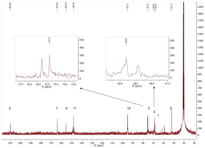
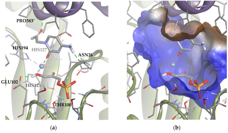
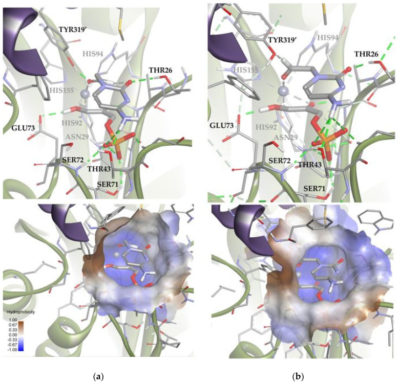
Dihydroxyacetone phosphate (DHAP)-dependent aldolases catalyze the aldol addition of DHAP to a variety of aldehydes and generate compounds with two stereocenters. This reaction is useful to synthesize chiral acyclic nucleosides, which constitute a well-known class of antiviral drugs currently used. In such compounds, the chirality of the aliphatic chain, which mimics the open pentose residue, is crucial for activity. In this work, three DHAP-dependent aldolases: fructose-1,6-biphosphate aldolase from rabbit muscle, rhanmulose-1-phosphate aldolase from Thermotoga maritima, and fuculose-1-phosphate aldolase from Escherichia coli, were used as biocatalysts. Aldehyde derivatives of thymine and cytosine were used as acceptor substrates, generating new acyclic nucleoside analogues containing two new stereocenters with conversion yields between 70% and 90%. Moreover, structural analyses by molecular docking were carried out to gain insights into the diasteromeric excess observed.

Keywords: aldol reaction; biocatalysis; drug design; stereoselectivity.


Conflict of interest statement

The authors declare no conflicts of interest.

Figures

Figure 1
Figure 1
Representative acyclic nucleosides with medicinal activities.
Figure 2
Figure 2
Stereoselective products generated by the four DHAP-dependent aldolases.
Figure 3
Figure 3
Expected acyclic nucleoside analogues produced by three different aldolases.
Figure 4
Figure 4
Partial view of the 1H NMR spectrum of the aldol product (2a+3a) obtained by RAMA bio-catalyzed aldol addition with thyminyl acetaldehyde and DHAP as substrates. The inset shows the different coupling constants of C3 hydrogens (J3–4 = 1.9 Hz) and C3′ hydrogens (J3′–4′ = 6.9 Hz), indicating 2a and 3a presence, respectively.
Figure 5
Figure 5
Coupling constants between neighboring protons [67].
Figure 6
Figure 6
Partial view of the 1H NMR spectrum of the aldol product (4b+5b) obtained by TmRhua-1PA bio-catalyzed aldol addition, with 1b and DHAP as substrates. The inset shows C3 hydrogen (J3–4 = 1.9 Hz) and C3′ hydrogen (J3′–4′ = 4.7 Hz) signals, indicating 4b and 5b presence, respectively.
Figure 7
Figure 7
Partial view of 13C NMR spectrum of the aldol products (4b+5b) obtained byTmRhua-1PA bio-catalyzed aldol addition and 1b as a substrate. The insets show the double signals corresponding to the chiral carbons C3 and C4, confirming the presence of both 4b and 5b isomers.
Figure 8
Figure 8
Partial view of 1H NMR spectrum of the aldol product (4b+5b) obtained by EcFuc-1PA bio-catalyzed aldol addition of 1b and DHAP as substrates. The inset shows the C3 (J3–4 = 1.6 Hz) and C3′ (J3′–4′ = 5.5 Hz) hydrogen signals.
Figure 9
Figure 9
Comparison between the active sites of two DHAP-dependent aldolases: (a) DHAP anchored in the EcFuc-1PA active site and (b) DHAP anchored in the TmRhu-1PA active site. Hydrophobicity is mapped onto a Connolly solvent-accessible surface of the receptor. Non-polar hydrogen atoms are omitted for clarity. Atom color code: C (grey), N (blue), O (red), Zn/Co (violet), P (orange), and H (white). The two protein chains are depicted with different colors (green and turquoise).
Figure 10
Figure 10
Docking for the cytosine derivative 1b with TmRhu-1PA, generating Solution 1: (a) DHAP coordinated by the carbonyl group of 1b ring, and (b) the -NH2 group of 1b interacting with the lateral hydrophilic pocket and the aldehyde group of the derivate with the upper hydrophilic pocket of the active site. The interactions are represented as dashed lines: green (H-bonds) and grey (coordination bonds). In (b), hydrophobicity is mapped onto a Connolly solvent-accessible surface of the receptor. Non-polar hydrogen atoms are omitted for clarity. Atom color code: C (grey), N (blue), O (red), Zn/Co (violet), P (orange), and H (white). The two protein chains are depicted with different colors (green and turquoise).
Figure 11
Figure 11
Docking for the cytosine derivative 1b with TmRhu-1PA and DHAP generating two possible solutions: (a) Solution 2: the R group is directed toward the upper hydrophilic pocket and (b) Solution 3: the R group is directed toward the lower hydrophilic pocket. The interactions are represented as dashed lines: green (H-bonds), grey (coordination bonds), and pink (pi-mediated interactions). Hydrophobicity is mapped onto a Connolly solvent-accessible surface of the receptor. Non-polar hydrogen atoms are omitted for clarity. Atom color code: C (grey), N (blue), O (red), Co (violet), P (orange), and H (white). The two protein chains are depicted with different colors (green and turquoise).
Figure 11
Figure 11
Docking for the cytosine derivative 1b with TmRhu-1PA and DHAP generating two possible solutions: (a) Solution 2: the R group is directed toward the upper hydrophilic pocket and (b) Solution 3: the R group is directed toward the lower hydrophilic pocket. The interactions are represented as dashed lines: green (H-bonds), grey (coordination bonds), and pink (pi-mediated interactions). Hydrophobicity is mapped onto a Connolly solvent-accessible surface of the receptor. Non-polar hydrogen atoms are omitted for clarity. Atom color code: C (grey), N (blue), O (red), Co (violet), P (orange), and H (white). The two protein chains are depicted with different colors (green and turquoise).
Figure 12
Figure 12
Docking for the thymine derivative 1a with TmRhu-1PA: (a) Solution 4: the aldehydic hydrogen shifts its orientation, exposing the re face to the DHAP. (b) Solution 5: the aldehydic hydrogen exposes the si face to the DHAP. The interactions are represented as dashed lines: green (H-bonds), light green (non-conventional H-bonds), grey (coordination bonds), and pink (pi-mediated interactions). Hydrophobicity is mapped onto a Connolly solvent-accessible surface of the receptor. Non-polar hydrogen atoms are omitted for clarity. Atom color code: C (grey), N (blue), O (red), Zn/Co (violet), P (orange), and H (white). The two protein chains are depicted with different colors (green and turquoise).
Figure 13
Figure 13
Docking for the cytosine derivative 1b attached by the aldehyde group to the metal ion of the active site of EcFuc-1PA: (a) Solution 6: the aldehydic hydrogen shifts its orientation, exposing the re face to the DHAP. (b) Solution 7: the aldehydic hydrogen exposes the si face to the DHAP. The interactions are represented as dashed lines: green (H-bonds), light green (non-conventional H-bonds), grey (coordination bonds), and pink (pi-mediated interactions). Hydrophobicity is mapped onto a Connolly solvent-accessible surface of the receptor. Non-polar hydrogen atoms are omitted for clarity. Atom color code: C (grey), N (blue), O (red), Zn/Co (violet), P (orange), and H (white). The two protein chains are depicted with different colors (green and turquoise).
Figure 14
Figure 14
Docking for the thymine derivative 1a attached by the aldehyde group to the metal ion of the active site of EcFuc-1PA: (a) Solution 8: Van der Waals interactions optimized in the hydrophobic pocket of the active site. (b) Solution 9: increase in the intensity of the substrate 1a–protein hydrogen bonds while minimizing the steric clash. The interactions are represented as dashed lines: green (H-bonds), light green (non-conventional H-bonds), grey (coordination bonds), and pink (pi-mediated interactions). Hydrophobicity is mapped onto a Connolly solvent-accessible surface of the receptor. Non-polar hydrogen atoms are omitted for clarity. Atom color code: C (grey), N (blue), O (red), Zn/Co (violet), P (orange), and H (white). The two protein chains are depicted with different colors (green and turquoise).

Similar articles

References

    1. Geraghty R.J., Aliota M.T., Bonnac L.F. Broad-Spectrum Antiviral Strategies and Nucleoside Analogues. Viruses. 2021;13:667. doi: 10.3390/v13040667. - DOI - PMC - PubMed
    1. Mirza A.Z. Advancement in the Development of Heterocyclic Nucleosides for the Treatment of Cancer—A Review. Nucleosides Nucleotides Nucleic Acids. 2019;38:836–857. doi: 10.1080/15257770.2019.1615623. - DOI - PubMed
    1. De Clercq E., Li G. Approved Antiviral Drugs over the Past 50 Years. Clin. Microbiol. Rev. 2016;29:695–747. doi: 10.1128/CMR.00102-15. - DOI - PMC - PubMed
    1. Eriksson U., Peterson L.W., Kashemirov B.A., Hilfinger J.M., Drach J.C., Borysko K.Z., Breitenbach J.M., Kim J.S., Mitchell S., Kijek P., et al. Serine Peptide Phosphoester Prodrugs of Cyclic Cidofovir: Synthesis, Transport, and Antiviral Activity. Mol. Pharm. 2008;5:598–609. doi: 10.1021/mp8000099. - DOI - PMC - PubMed
    1. Gómez-Coca R.B., Blindauer C.A., Sigel A., Operschall B.P., Holý A., Sigel H. Extent of Intramolecular π-Stacks in Aqueous Solution in Mixed-Ligand Copper(II) Complexes Formed by Heteroaromatic Amines and Several 2-Aminopurine Derivatives of the Antivirally Active Nucleotide Analog 9-[2-(Phosphonomethoxy) Ethyl]Adenine (PMEA) Chem. Biodivers. 2012;9:2008–2034. doi: 10.1002/cbdv.201200022. - DOI - PubMed

MeSH terms

Substances

LinkOut - more resources