. 2024 Jun 27;14(7):765.
doi: 10.3390/biom14070765.

Biochemical and Functional Profiling of Thioredoxin-Dependent Cytosolic GPX-like Proteins in Euglena gracilis

Affiliations

Biochemical and Functional Profiling of Thioredoxin-Dependent Cytosolic GPX-like Proteins in Euglena gracilis

Md Topu Raihan et al. Biomolecules. .

Abstract

Unlike plants and animals, the phytoflagellate Euglena gracilis lacks catalase and contains a non-selenocysteine glutathione peroxidase-like protein (EgGPXL), two peroxiredoxins (EgPrx1 and EgPrx4), and one ascorbate peroxidase in the cytosol to maintain reactive oxygen species (ROS) homeostasis. In the present study, the full-length cDNA of three cytosolic EgGPXLs was obtained and further characterized biochemically and functionally. These EgGPXLs used thioredoxin instead of glutathione as an electron donor to reduce the levels of H2O2 and t-BOOH. The specific peroxidase activities of these enzymes for H2O2 and t-BOOH were 1.3 to 4.9 and 0.79 to 3.5 µmol/min/mg protein, respectively. Cytosolic EgGPXLs and EgPrx1/EgPrx4 were silenced simultaneously to investigate the synergistic effects of these genes on the physiological function of E. gracilis. The suppression of cytosolic EgGPXL genes was unable to induce any critical phenomena in Euglena under normal (100 μmol photons m-2 s-1) and high-light conditions (350 μmol photons m-2 s-1) at both autotrophic and heterotrophic states. Unexpectedly, the suppression of EgGPXL genes was able to rescue the EgPrx1/EgPrx4-silenced cell line from a critical situation. This study explored the potential resilience of Euglena to ROS, even with restriction of the cytosolic antioxidant system, indicating the involvement of some compensatory mechanisms.

Keywords: Euglena gracilis; cytosolic GPX-like protein; reactive oxygen species.


Conflict of interest statement

The authors declare no conflict of interest.

Figures

Figure 1
Figure 1
Sequence analysis of GPXL proteins derived from Euglena as well as other organisms. Three GPX signature motifs were found ubiquitously across all domains of GPXLs distinguished by green boxes. A basic (Asp), a nonpolar (Try), and a thiol (Cys) amino acid, denoted by green triangle, might be responsible for catalysis, highly conserved in almost all the GPXLs from various domains. In contrast, plant GPXs and EgGPXLs contained two cysteine residues, which were indicated in orange triangle and green triangle. Additionally, amino acid residues demonstrating identical characteristics throughout all sequences were marked with a red asterisk below. Furthermore, residues displaying similar traits were indicated with paired dots beneath the sequences, whereas those with a degree of resemblance were identified with a single dot. EgGPXLs were highlighted with blue color. Selenocysteine (U), found in many prokaryotes and eukaryotes except plants, certain insects, nematodes, and various protists, was replaced evolutionarily by cysteine in remaining GPXs (highlighted in yellow color).
Figure 2
Figure 2
SDS-PAGE-based expression and purification analysis of 6X-His tagged rEgGPXL-2 (A) and rEgGPXL-4 (B). The rEgGPXL-2 and -4 were taken in TALON Metal Affinity resin for purification after expression in E. coli Bl21. Eight types of samples were run in this electrophoresis system which were input from Lane 1 to 7. Sample after expression, flow-through (first fraction in purification), sample obtained in first washing step, and sample obtained after last washing step were loaded in Lanes 2 to 4, correspondingly. Through three consecutive elutions loaded in Lanes 5 to 7, respectively, we tried to find purified proteins. After separation of proteins through SDS/PAGE, it was visualized with Coomassie Brilliant Blue. M denoted the molecular weight marker.
Figure 3
Figure 3
The kinetic behavior of cytosolic rEgGPXLs. Hyperbolic graph was obtained for rEgGPXL-2 and rEgGPXL-4 when H2O2 (A,B) and t-BOOH (C,D)-dependent peroxidase activity was performed for these cytosolic proteins, respectively. (A,B) denoted H2O2 and t-BOOH-dependent peroxidase activity for rEgGPXL-2, respectively; similarly, (C,D) denoted it for rEgGPXL-4. The Lineweaver–Burk plots (inset) were constructed through best fit lines. The values represented the mean average of three consecutive experimental runs.
Figure 4
Figure 4
Thioredoxin (Trx)-dependent peroxidase activity of cytosolic rEgGPXLs. (A,B) indicated Trx-dependent peroxidase activity using H2O2 and t-BOOH as substrates for rEgGPXL-2, respectively. Similarly, (C,D) denoted it for rEgGPXL-4. The values represented the mean average of three consecutive experimental runs.
Figure 5
Figure 5
pH dependency of cytosolic rEgGPXLs. This result implied optimum activity of rEgGPXL-2 (A) and rEgGPXL-4 (B) from slightly acidic to neutral pH. After pH 8.0 and above, a significant reduction of peroxidase activity was noted in both enzymes. The values represented the mean average of three consecutive experimental runs.
Figure 6
Figure 6
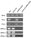
RT-PCR-based confirmation of cytosolic EgGPXLs silencing. In addition to cytosolic EgGPXLs, another two cytosolic peroxidases, EgPrx-1 and EgPrx-4, were also silenced for investigation of synergistic effect of these cytosolic peroxidases. Therefore, quantitative analysis of gene expression was conducted for five types of peroxidase genes named EgGPXL-2, EgGPXL-3, EgGPXL-4, EgPrx-1, EgPrx-4, and one housekeeping gene, EF1α, through RT-PCR. PCR amplification of EgGPXL-2, EgGPXL-3, EgGPXL-4, EgPrx-1, EgPrx-4, and EF1α was conducted using cDNA templates obtained from the WT and mutant cell lines. Lane 1 contained WT (control, electroporation without dsRNA); Lanes 2 to 4 contained samples from three mutant cell lines, ΔPrx-1/4, ΔGPXL-2/-3/4, and ΔPrx-1/4, GPXL-2/3/4, respectively. The result was confirmed after repeating three consecutive experimental runs.
Figure 7
Figure 7
Observation of phenotypic change after suppression of cytosolic EgGPXLs. In addition to cytosolic EgGPXLs, another two cytosolic peroxidases, EgPrx-1 and EgPrx-4, were also silenced for investigation of synergistic effect of these cytosolic peroxidases. Wild-type and mutant cell lines were applied as spots onto KH plates used for heterotrophic growth conditions (A,B) and onto CM plates designated for autotrophic growth conditions (C,D). The cell quantities spotted were 6 × 10⁶, 3 × 10⁶, 15 × 105, 7.5 × 10⁵, and 3.75 × 10⁵ in both agar plates. Subsequently, the plates were taken to a 7-day incubation period under normal growth conditions (26 °C with 100 μmol photons m−2 s−1) (A,C) and high-light conditions (300 μmol photons m−2 s−1) (B,D) to induce oxidative stress in the cell lines. ΔPrx-1/4, ΔGPXL-2/3/4, and ΔPrx-1/4, GPXL-2/3/4 corresponded to the cell lines where Prx-1/Prx-4, GPXL-2/GPXL-3/GPXL-4, and Prx-1/Prx-4, GPXL-2/GPXL-3/GPXL-4 genes were suppressed, respectively. The result was confirmed after performing three consecutive replications.
Figure 8
Figure 8
Growth rates of the control and each KD cell. Cells were grown heterotrophically under dark at 26 °C for 8 days. ΔPrx-1/4, ΔGPXL-2/3/4, and ΔPrx-1/4, GPXL-2/3/4 corresponded to the cell lines where Prx-1/Prx-4, GPXL-2/GPXL-3/GPXL-4, and Prx-1/Prx-4, GPXL-2/GPXL-3/GPXL-4 genes were suppressed, respectively. Values are the mean ± SD (n = 3). Values with asterisks were significantly different, according to the ANOVA (p < 0.05).
Figure 9
Figure 9
Effect of suppression of EgGPXLs on APX and Trx dependent peroxidase activity in the wild-type (WT) and mutant cell lines. APX (A) and Trx-dependent peroxidase (B) were checked in extracts derived from both the wild-type cells and each individual mutant cell line, respectively. ΔPrx-1/4, ΔGPXL-2/3/4, and ΔPrx-1/4, GPXL-2/3/4 corresponded to the cell lines where Prx-1/Prx-4, GPXL-2/GPXL-3/GPXL-4, and Prx-1/Prx-4, GPXL-2/GPXL-3/GPXL-4 genes were suppressed, respectively. Values are the mean ± SD (n = 3). Values with same letter indicated no significant difference in enzyme activity, while those with different letters had significant differences in enzyme activity in all cell lines according to Dunnet t-test (p < 0.05).
Figure 10
Figure 10
Assessment of chlorophyll content in mutants and wild-type cell line of Euglena. ΔPrx-1/4, ΔGPXL-2/3/4, and ΔPrx-1/4, GPXL-2/3/4 corresponded to the cell lines where Prx-1/Prx-4, GPXL-2/GPXL-3/GPXL-4, and Prx-1/Prx-4, GPXL-2/GPXL-3/GPXL-4 genes were suppressed, respectively. Chlorophyll was measured for both chlorophyll a and b, according to previously mentioned protocol, without further modification [32]. Values with same letter indicated no significant difference in chlorophyll content in mutant cell lines compared to wild type, according to Dunnet t-test (p < 0.05).
Figure 11
Figure 11
Quantitative measurement of glutathione (A) and its redox ratio (B) in mutants and wild-type cell lines of Euglena. ΔPrx-1/4, ΔGPXL-2/3/4, and ΔPrx-1/4, GPXL-2/3/4 corresponded to the cell lines where Prx-1/Prx-4, GPXL-2/GPXL-3/GPXL-4, and Prx-1/Prx-4, GPXL-2/GPXL-3/GPXL-4 genes were suppressed, respectively. Total, reduced, and oxidized glutathione were measured, and no significant difference was observed in any cell lines compared to the wild type. Values with the same letter indicated no significant difference in glutathione content in mutant cell lines compared to the wild-type, according to Dunnet t-test (p < 0.05).
Figure 12
Figure 12
H2O2 quantitation in mutants and wild-type cell lines of Euglena. ΔPrx-1/4, ΔGPXL-2/3/4, and ΔPrx-1/4, GPXL-2/3/4 corresponded to the cell lines where Prx-1/Prx-4, GPXL-2/GPXL-3/GPXL-4, and Prx-1/Prx-4, GPXL-2/GPXL-3/GPXL-4 genes were suppressed, respectively. Flurointensity among all the cell lines was determined by fluorescence detection system (Corona-SH 9000) using H2O2-specific fluorescent probe known as BES- H2O2-Ac (Derivative of BES (benzenesulfonyl) (Wako, Japan)). Values with same letter indicated no significant difference in H2O2 content in mutant cell line compared to wild type, according to Dunnet t-test (p < 0.05).

Similar articles

References

    1. Mittler R., Zandalinas S.I., Fichman Y., Van Breusegem F. Reactive Oxygen Species Signalling in Plant Stress Responses. Nat. Rev. Mol. Cell Biol. 2022;23:663–679. doi: 10.1038/s41580-022-00499-2. - DOI - PubMed
    1. Waszczak C., Akter S., Jacques S., Huang J., Messens J., Van Breusegem F. Oxidative Post-Translational Modifications of Cysteine Residues in Plant Signal Transduction. J. Exp. Bot. 2015;66:2923–2934. doi: 10.1093/jxb/erv084. - DOI - PubMed
    1. Medrano-Macías J., Flores-Gallegos A.C., Nava-Reyna E., Morales I., Tortella G., Solís-Gaona S., Benavides-Mendoza A. Reactive Oxygen, Nitrogen, and Sulfur Species (RONSS) as a Metabolic Cluster for Signaling and Biostimulation of Plants: An Overview. Plants. 2022;23:3203. doi: 10.3390/plants11233203. - DOI - PMC - PubMed
    1. Manta B., Comini M., Medeiros A., Hugo M., Trujillo M., Radi R. Trypanothione: A Unique Bis-Glutathionyl Derivative in Trypanosomatids. Biochim. Biophys. Acta. 2013;1830:3199–3216. doi: 10.1016/j.bbagen.2013.01.013. - DOI - PubMed
    1. Navrot N., Collin V., Gualberto J., Gelhaye E., Hirasawa M., Rey P., Knaff D.B., Issakidis E., Jacquot J.P., Rouhier N. Plant Glutathione Peroxidases are Functional Peroxiredoxins Distributed in Several Subcellular Compartments and Regulated During Biotic and Abiotic Stresses. Plant Physiol. 2006;142:1364–1379. doi: 10.1104/pp.106.089458. - DOI - PMC - PubMed

MeSH terms

LinkOut - more resources