. 2024 Jun 28;14(7):772.
doi: 10.3390/biom14070772.

Helicobacter pylori-Induced Decrease in Membrane Expression of Na,K-ATPase Leads to Gastric Injury

Affiliations

Helicobacter pylori-Induced Decrease in Membrane Expression of Na,K-ATPase Leads to Gastric Injury

Olga Vagin et al. Biomolecules. .

Abstract

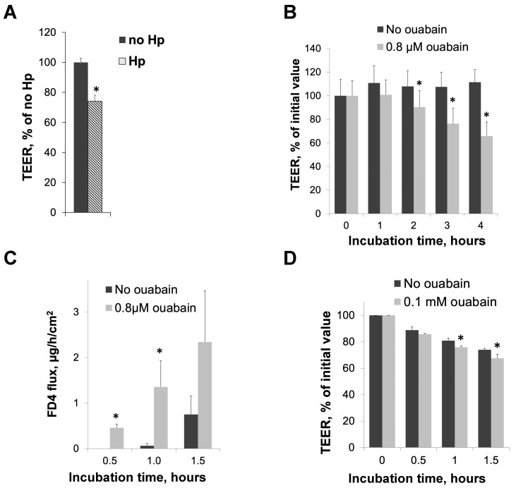
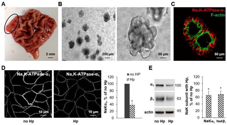
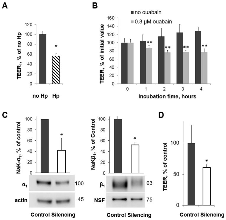
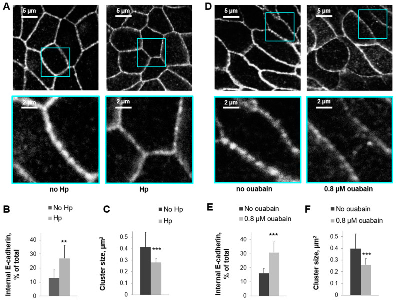
Helicobacter pylori is a highly prevalent human gastric pathogen that causes gastritis, ulcer disease, and gastric cancer. It is not yet fully understood how H. pylori injures the gastric epithelium. The Na,K-ATPase, an essential transporter found in virtually all mammalian cells, has been shown to be important for maintaining the barrier function of lung and kidney epithelia. H. pylori decreases levels of Na,K-ATPase in the plasma membrane of gastric epithelial cells, and the aim of this study was to demonstrate that this reduction led to gastric injury by impairing the epithelial barrier. Similar to H. pylori infection, the inhibition of Na,K-ATPase with ouabain decreased transepithelial electrical resistance and increased paracellular permeability in cell monolayers of human gastric cultured cells, 2D human gastric organoids, and gastric epithelium isolated from gerbils. Similar effects were caused by a partial shRNA silencing of Na,K-ATPase in human gastric organoids. Both H. pylori infection and ouabain exposure disrupted organization of adherens junctions in human gastric epithelia as demonstrated by E-cadherin immunofluorescence. Functional and structural impairment of epithelial integrity with a decrease in Na,K-ATPase amount or activity provides evidence that the H. pylori-induced downregulation of Na,K-ATPase plays a role in the complex mechanism of gastric disease induced by the bacteria.

Keywords: Helicobacter pylori; Na,K-ATPase; adherens junctions; gastric injury; ouabain.


Conflict of interest statement

The authors report no conflicts of interest.

Figures

Figure 1
Figure 1
Both Helicobacter pylori infection and reduced Na,K-ATPase activity impair intracellular junctions in cultured cells (AC) and in gerbil mucosa (D). HGE-20 cells were grown to confluency in Transwell™ inserts, and the transepithelial electrical resistance (TEER) was measured in the presence of H. pylori (A) or ouabain (B). The TEER is expressed as a percentage of the initial value. Initial values of the TEER in HGE-20 cells ranged from 1000 to 1500 Ω×cm2. Paracellular permeability of confluent HGE-20 cell layers was measured in an Ussing chamber in the presence or absence of ouabain, results are expressed as the rate of FD4 flux through the cell layer (C). Gerbils were euthanized and stomach tissue incubated in an Ussing chamber in the presence of ouabain, and the TEER was measured at the indicated times. Results are expressed as percent of initial value (D). Initial values of the TEER in gerbil gastric mucosa ranged from 75 to 100 Ω×cm2, consistent with prior published results [57]. For all panels, n = 3, * p < 0.05 (t-test), mean ± SD.
Figure 2
Figure 2
Human gastric organoids as a model system to study the effect of Helicobacter pylori on gastric mucosa. Human gastric antrum was obtained from de-identified sleeve gastrectomy specimens. The location of the harvested sample is indicated by the circle (A). Human gastric organoids were cultivated from the fresh tissue samples and plated in Matrigel with appropriate growth factors, leading to the formation of 3D organoids (B). Basolateral expression of Na,K-ATPase was confirmed in gastric organoids by immunofluorescence of the α1 subunit, indicated by the red color, and F-actin was used as a counterstain, indicated by the green color (C). Three-dimensional organoids were converted to 2D using Matrigel-coated Transwell™ inserts and were grown into a confluent monolayer. Organoids were incubated with the vehicle or H. pylori (D,E), and the decrease in Na,K-ATPase subunits in the presence of H. pylori was confirmed in this model system by immunofluorescence (D) or Western blot (E). Quantification is shown in the right panels. Mean ± SD, n = 3, * p < 0.05, t-test.
Figure 3
Figure 3
Helicobacter pylori infection, reduced amount of Na,K-ATPase, and reduced Na,K-ATPase activity similarly decrease the TEER in human gastric organoids. (A,B) The TEER of 2D human gastric organoids grown on Transwell™ inserts and measured in an Endohm® chamber was decreased after exposure to Helicobacter pylori (A) and in the presence of ouabain compared with the vehicle (B). Western blot showing the silencing of the Na,K-ATPase α1 subunit (C). The TEER of 2D human gastric organoids grown on Transwell™ inserts and measured in an Endohm® chamber was decreased after silencing the Na,K-ATPase α1 subunit (D). Initial values of the TEER in 2D gastric organoids ranged from 800 to 1300 Ω×cm2. Mean ± SD, significant differences from control, t-test, n = 3, * p < 0.05 (A,C,D) and n = 5, ** p < 0.01 (B).
Figure 4
Figure 4
Reduced Na,K-ATPase activity and Helicobacter infection similarly impair adherens junctions in human gastric organoids. Immunofluorescence of 2D gastric organoids using E-cadherin antibodies showed a decreased E-cadherin signal at the cell junctions in the presence of H. pylori (A) or ouabain (D) and an increased intracellular accumulation of E-cadherin (B,E). E-cadherin clusters were decreased in size in the presence of H. pylori (C) or ouabain (F), suggesting injury to junctions. Mean ± SD, significant differences from “no Hp” or “no ouabain”, t-test. n = 5, ** p < 0.01, *** p < 0.001 (B,F).

Similar articles

References

    1. Blaser M.J. Hypotheses on the pathogenesis and natural history of Helicobacter pylori-induced inflammation. Gastroenterology. 1992;102:720–727. doi: 10.1016/0016-5085(92)90126-J. - DOI - PubMed
    1. Nomura A., Stemmermann G.N., Chyou P.H., Kato I., Perez-Perez G.I., Blaser M.J. Helicobacter pylori infection and gastric carcinoma among Japanese Americans in Hawaii. N. Engl. J. Med. 1991;325:1132–1136. doi: 10.1056/NEJM199110173251604. - DOI - PubMed
    1. Parsonnet J. Gastric adenocarcinoma and Helicobacter pylori infection. West. J. Med. 1994;161:60. - PMC - PubMed
    1. Parsonnet J., Friedman G.D., Vandersteen D.P., Chang Y., Vogelman J.H., Orentreich N., Sibley R.K. Helicobacter pylori infection and the risk of gastric carcinoma. N. Engl. J. Med. 1991;325:1127–1131. doi: 10.1056/NEJM199110173251603. - DOI - PubMed
    1. Suerbaum S., Michetti P. Helicobacter pylori infection. N. Engl. J. Med. 2002;347:1175–1186. doi: 10.1056/NEJMra020542. - DOI - PubMed

MeSH terms