. 2024 Jun 29;14(7):779.
doi: 10.3390/biom14070779.

Anti-Biofilm Perspectives of Propolis against Staphylococcus epidermidis Infections

Affiliations

Anti-Biofilm Perspectives of Propolis against Staphylococcus epidermidis Infections

Virginia Vadillo-Rodríguez et al. Biomolecules. .

Abstract

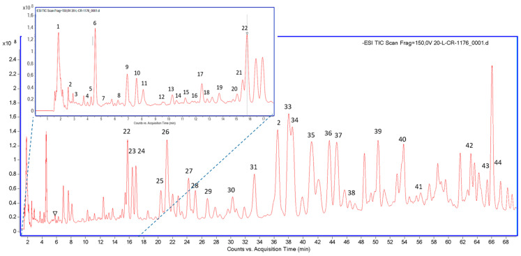
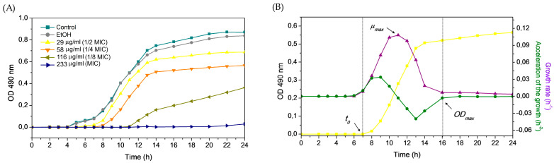
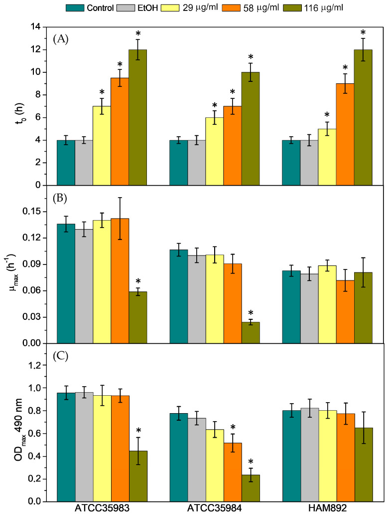
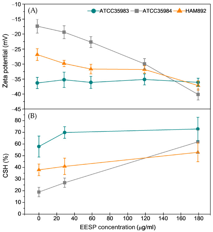
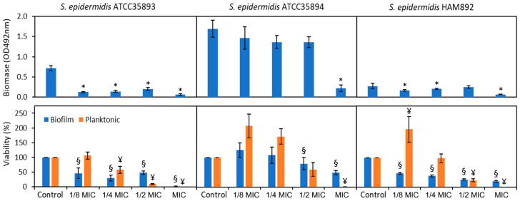
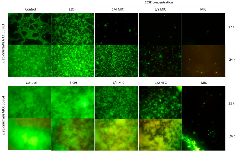
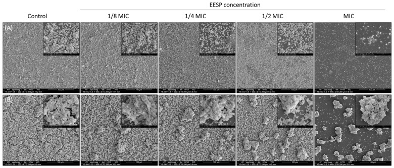
Staphylococcus epidermis has emerged as the main causative agent of medical device-related infections. Their major pathogenicity factor lies in its ability to adhere to surfaces and proliferate into biofilms, which increase their resistance to antibiotics. The main objective of this study was to evaluate the use and the mechanism of action of an ethanolic extract of Spanish propolis (EESP) as a potential alternative for preventing biofilm-related infections caused by S. epidermidis. The chemical composition of propolis is reported and its antibacterial activity against several strains of S. epidermidis with different biofilm-forming capacities evaluated. The influence of sub-inhibitory concentrations (sub-MICs) of EESP on their growth, physicochemical surface properties, adherence, and biofilm formation were studied. EESP interferes with planktonic cells, homogenizing their physicochemical surface properties and introducing a significant delay in their growth. The adherence and biofilms at the EESP concentrations investigated were decreased up to 90.5% among the strains. Microscopic analysis indicated that the planktonic cells that survived the treatment were the ones that adhere and proliferate on the surfaces. The results obtained suggest that the EESP has a high potential to be used as an inhibitor of both the adhesion and biofilm formation of S. epidermidis.

Keywords: Staphylococcus epidermidis; biofilms; hydrophobicity; propolis; zeta potential.


Conflict of interest statement

The authors declare no conflict of interest.

Figures

Figure 1
Figure 1
LC–MS chromatogram of EESP showing the major and minor organic peaks found in the sample.
Figure 2
Figure 2
(A) Growth curves obtained for S. epidermidis ATCC 35984 under the different concentrations of EESP studied. (B) Example of the growth curve recorded for this strain at the EESP concentration of 58 μg/mL (¼ of the MIC) and of its first and second derivative, i.e., growth rate and acceleration of the growth, respectively, from which the main growth parameters, i.e., lag time duration (t0), maximum growth rate (μmax) and final optical density (ODmax), were obtained.
Figure 3
Figure 3
(A) Lag phase duration (t0), (B) maximum growth rate (µmax) and (C) final optical density (ODmax) obtained from the analyses of the growth curves of the cells for the controls and the different sub-MIC concentrations of EESP investigated. Data shown as mean ± SD. * Statistically significant differences versus the control.
Figure 4
Figure 4
Zeta potential values (A) and degree of cell surface hydrophobicity assessed by MATH (B) measured for the strains investigated in the absence and presence of the different sub-MIC concentrations of EESP tested.
Figure 5
Figure 5
Biofilm biomass and viability of the adherent and planktonic cells determined for the three strains investigate in the absence and present of the different sub-MIC concentrations of EESP. Statistically significant differences versus the control (*, ¥, §).
Figure 6
Figure 6
Fluorescence microscopy images of the biofilms produced by S. epidermidis ATCC 35983 and ATCC 35984 under the different conditions investigated (i.e., controls and sub-MIC concentrations of EESP) after an incubation time of 12 and 24 h.
Figure 7
Figure 7
Scanning electron micrographs of the biofilms produced by (A) S. epidermidis ATCC 35983 and (B) ATCC 35984 under the different conditions investigated (i.e., controls and sub-MIC concentrations of EESP) after an incubation time of 24 h. Image magnification: 1000× and 10,000×.

Similar articles

Cited by

References

    1. Aslam B., Wang W., Arshad M.I., Khurshid M., Muzammil S., Rasool M.H., Nisar M.A., Alvi R.F., Aslam M.A., Qamar M.U., et al. Antibiotic resistance: A rundown of a global crisis. Infect. Drug. Resist. 2018;11:1645–1658. doi: 10.2147/IDR.S173867. - DOI - PMC - PubMed
    1. Khatoon Z., McTiernan C.D., Suuronen E.J., Mah T.F., Alarcon E.I. Bacterial biofilm formation on implantable devices and approaches to its treatment and prevention. Heliyon. 2018;4:e01067. doi: 10.1016/j.heliyon.2018.e01067. - DOI - PMC - PubMed
    1. Pontes J.T.C., Toledo-Borges A.B., Roque-Borda C.A., Pavan F.R. Antimicrobial peptides as an alternative for the eradication of bacterial biofilms of multi-drug resistant bacteria. Pharmaceutics. 2022;15:642. doi: 10.3390/pharmaceutics14030642. - DOI - PMC - PubMed
    1. Wang N., Luo J., Deng F., Huang Y., Zhou H. Antibiotic combination therapy: A strategy to overcome bacterial resistance to aminoglycoside antibiotics. Front. Pharmacol. 2022;23:839808. doi: 10.3389/fphar.2022.839808. - DOI - PMC - PubMed
    1. Mhlongo J.T., Waddad A.Y., Albericio F., de la Torre B.G. Antimicrobial peptide synergies for fighting infectious diseases. Adv. Sci. 2023;10:2300472. doi: 10.1002/advs.202300472. - DOI - PMC - PubMed

MeSH terms

LinkOut - more resources