Review
. 2024 Jul 14;14(7):848.
doi: 10.3390/biom14070848.

Role of Type I Interferons during Mycobacterium tuberculosis and HIV Infections

Affiliations
Review

Role of Type I Interferons during Mycobacterium tuberculosis and HIV Infections

Elsa Anes et al. Biomolecules. .

Abstract

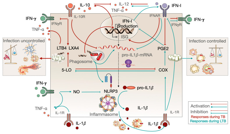
Tuberculosis and AIDS remain two of the most relevant human infectious diseases. The pathogens that cause them, Mycobacterium tuberculosis (Mtb) and HIV, individually elicit an immune response that treads the line between beneficial and detrimental to the host. Co-infection further complexifies this response since the different cytokines acting on one infection might facilitate the dissemination of the other. In these responses, the role of type I interferons is often associated with antiviral mechanisms, while for bacteria such as Mtb, their importance and clinical relevance as a suitable target for manipulation are more controversial. In this article, we review the recent knowledge on how these interferons play distinct roles and sometimes have opposite consequences depending on the stage of the pathogenesis. We highlight the dichotomy between the acute and chronic infections displayed by both infections and how type I interferons contribute to an initial control of each infection individually, while their chronic induction, particularly during HIV infection, might facilitate Mtb primo-infection and progression to disease. We expect that further findings and their systematization will allow the definition of windows of opportunity for interferon manipulation according to the stage of infection, contributing to pathogen clearance and control of immunopathology.

Keywords: HIV; co-infection; interferons; tuberculosis.


Conflict of interest statement

The authors declare no conflicts of interest.

Figures

Figure 1
Figure 1
The evolution of vertebrates from jawless to jawed species occurred concurrently with two rounds of whole-genome duplications. The first (star labeled 1R) and the second (star labeled 2R) are estimated to have occurred approximately 450 million years ago. This genome expansion resulted in the incorporations of cassettes of adaptive immunity, along with cytokines and their receptors. One hypothesis suggests that IFNs originated within the class II α-helical cytokine family, where antiviral interferons form a clade along with the interleukin (IL)-10 cytokine family. All three IFN classes were present in the jawed vertebrate ancestor.
Figure 2
Figure 2
Schematic representation of innate pattern recognition receptors (PRRS) and the subsequent activation of IFN and ISGs. Cytoplasmic or endosomal membrane-associated toll-like receptors (TLRs) or cytosolic-associated immune sensors that recognize pathogen-associated molecular patterns (PAMPs) initiate a signaling cascade that results in the secretion of IFN, ISGs, and proinflammatory cytokines. Gamma interferon-inducible protein 16 (IFI16), DNA-dependent activator of IFN regulatory factors (DAI), and cyclic GMP-AMP synthase (cGas) are sensors for DNA. Retinoic acid-inducible gene 1 (RIG-I), melanoma differentiation-associated protein 5 (MDA5), and oligoadenylate synthetase (OAS) and latent endoribonuclease (RNaseL) recognize foreign RNA. The signaling proceeds to transcription factor activity by stimulator of IFN genes (STING) and mitochondrial antiviral-signaling protein (MAVS) at the ER/mitochondrion interface. Consequently, this results in the activation of interferon (IFN) response factors 3 or 7 (IRF3/7), or alternatively, the activation of NF-κB. These transcription factors translocate to the nucleus, where they activate specific promoters, triggering the expression of IFN and a first subset of ISGs. IFN is secreted out of the cell and signals through IFNAR, which is then transmitted to signal transducers and activators of transcription (STATS). This results in the expression of a large spectrum of ISGs with distinct functions, including antiviral/antimicrobial effectors, and negative (repressors of inflammation) or positive (inducers of inflammatory responses) regulators of IFN signaling. The disabling of the interferon responses may be induced by ISGs, such as the suppressor of cytokine signaling (SOCS) or ubiquitin carboxy-terminal hydrolase 18 (USP18), which block the signals directly from IFNAR or TLRs. In addition, they function as transcriptional repressors of proinflammatory cytokine transcription factors, such as C-Rel and IFIT1. STAT3 may act as a transcriptional repressor by sequestering STAT1. Secreted ISG15 stimulates an increase in the expression of immunosuppressive cytokine IL-10 and the programmed cell death ligand 1 (PD-L1) in monocytes. The equilibrium of these pathways may result in either protective or detrimental effects on the host. Gamma-activated sequences (GASs); interferon-stimulated response element (ISRE); IFN-inducible transmembrane (IFITM); IFN-induced protein with tetratricopeptide repeats (IFIT); virus inhibitory protein, endoplasmic reticulum-associated, IFN-inducible (Viperin); myxovirus resistance (Mx); cholesterol-25-hydroxylase (CH25H); tripartite motif protein 5α (TRIM5α); apolipoprotein B mRNA-editing enzyme catalytic polypeptide 3 (APOBEC3); HD domain-containing deoxynucleoside triphosphate triphosphohydrolase (SAMHD1).
Figure 3
Figure 3
Interferon-mediated antiviral mechanisms against HIV infection affect all stages of the viral cycle, from fusion, uncoating, reverse transcription, nuclear import, and translation, to virus particle release. HIV developed evasion mechanisms towards these IFN-I-induced restriction factors. APOBEC: apolipoprotein B mRNA-editing enzyme, catalytic polypeptide-like; IFITM: interferon-induced transmembrane protein; MX2: myxovirus resistance protein 2 (also known as MX dynamin like GTPase 2); SAMHD1: sterile alpha motif [SAM] and histidine/aspartic acid [HD] domain-containing protein 1; TRIM5α: tripartite motif 5 alpha.
Figure 4
Figure 4
Role of IFN-I during infection with Mtb. Basal levels of interferon signaling are protective for the host. Signaling via the IFNAR receptor results in the expression of hundreds of ISGs. Among these are the protective cytokines IL-12 and TNFα. The latter protects the host in an autocrine or paracrine manner, leading to the activation of the infected macrophage to a more bactericidal state. IFN-γ produced from IL-12-stimulated lymphocytes further increases the oxidative burst. Itaconate, produced by the immunoresponsive gene 1 (IRG1) from cis-aconitate, modulates the TCA cycle by regulating succinate dehydrogenase activity for fumarate production. During Mtb infection, first, itaconate induces mtROS and inducible nitric oxide synthase (iNOS), leading to pathogen control. Itaconate has additional antimicrobial activity that inhibits methyl citrate lyase (MCL) in the methyl citrate cycle (MCC) and isocitrate lyase (ICL) in the glyoxylate shunt, enzymes that are essential for Mtb survival. Moreover, itaconyl-coenzyme A (CoA) targets B12-dependent methylmalonyl-CoA mutase (MCM-B12), thereby inhibiting bacterial growth. High IFN-I signaling is observed during the progression of tuberculosis, with detrimental effects on the host. These effects are mainly due to the increased secretion of the immunosuppressor IL-10, a decreased secretion of protective cytokines, and opposing the protective effects of IFN-γ. This environment of chemoattractant signals attracts myeloid cells to the granuloma, which allows an uncontrolled bacterial replication concomitant with high levels of inflammatory cell death.
Figure 5
Figure 5
Immune responses during LTB and during progression to TB. Red lines represent the responses during active disease with high IFN-I signature signaling. This leads to the inhibition of IL-1β secretion, uncontrolled bacilli replication, and strong inflammatory responses through massive neutrophil recruitment and NETosis, which is followed by the stimulation of 5-LO products. These mechanisms culminate in lung cavitation and the eventual death of the patient in the absence of antibiotic treatment. The green lines represent the events that occur during LTB, which promote infection control through a series of phagocytic events and apoptotic cell death mediated by the stimulation of COX products; in contrast, IL-1β secretion is kept under protective, balanced levels.

Similar articles

Cited by

References

    1. Isaacs A., Lindenmann J. Virus interference. I. The interferon. Proc. R. Soc. Lond. B-Biol. Sci. 1957;147:258–267. doi: 10.1098/rspb.1957.0048. - DOI - PubMed
    1. Lindenmann J., Burke D.C., Isaacs A. Studies on the production, mode of action and properties of interferon. Br. J. Exp. Pathol. 1957;38:551–562. - PMC - PubMed
    1. Nagano Y., Kojima Y., Sawai Y. Immunity and interference in vaccinia; inhibition of skin infection by inactivated virus. Comptes Rendus Seances Soc. Biol. Fil. 1954;148:750–752. - PubMed
    1. Biondo C., Midiri A., Gambuzza M., Gerace E., Falduto M., Galbo R., Bellantoni A., Beninati C., Teti G., Leanderson T., et al. IFN-α/β Signaling Is Required for Polarization of Cytokine Responses toward a Protective Type 1 Pattern during Experimental Cryptococcosis. J. Immunol. 2008;181:566–573. doi: 10.4049/jimmunol.181.1.566. - DOI - PubMed
    1. Ishihara T., Aga M., Hino K., Ushio C., Taniguchi M., Iwaki K., Ikeda M., Kurimoto M. Inhibition of Chlamydia trachomatis growth by human interferon-alpha;: Mechanisms and synergistic effect with interferon-gamma; and tumor necrosis factor-alpha. Biomed. Res. 2005;26:179–185. doi: 10.2220/biomedres.26.179. - DOI - PubMed

Substances

LinkOut - more resources