Review
. 2024 Jul 19;14(7):866.
doi: 10.3390/biom14070866.

Calreticulin-Enigmatic Discovery

Affiliations
Review

Calreticulin-Enigmatic Discovery

Gillian C Okura et al. Biomolecules. .

Abstract

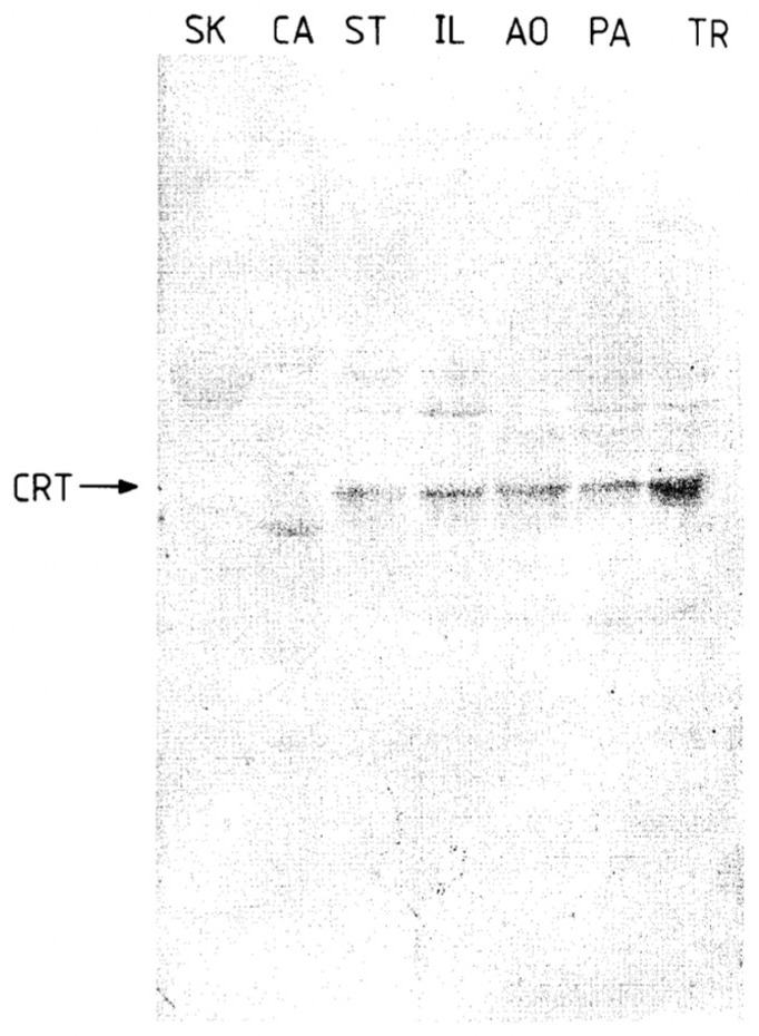
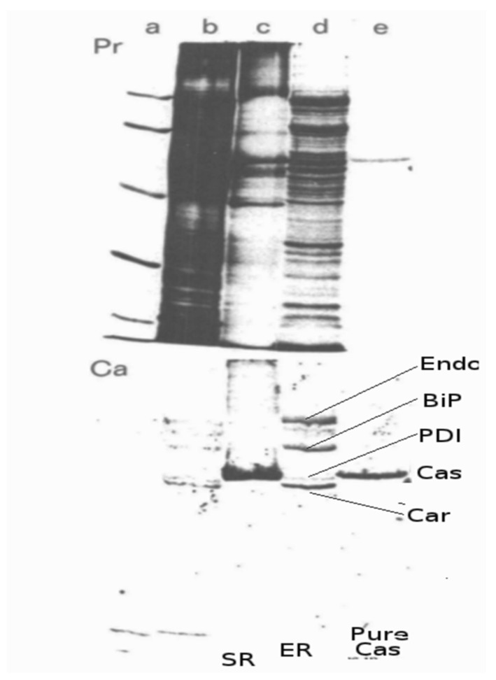
Calreticulin (CRT) is an intrinsically disordered multifunctional protein that plays essential roles intra-and extra-cellularly. The Michalak laboratory has proposed that CRT was initially identified in 1974 by the MacLennan laboratory as the high-affinity Ca2+-binding protein (HACBP) of the sarcoplasmic reticulin (SR). This widely accepted belief has been ingrained in the scientific literature but has never been rigorously tested. In our report, we have undertaken a comprehensive reexamination of this assumption by meticulously examining the majority of published studies that present a proteomic analysis of the SR. These analyses have utilized proteomic analysis of purified SR preparations or purified components of the SR, namely the longitudinal tubules and junctional terminal cisternae. These studies have consistently failed to detect the HACBP or CRT in skeletal muscle SR. We propose that the existence of the HACBP has failed the test of reproducibility and should be retired to the annals of antiquity. Therefore, the scientific dogma that the HACBP and CRT are identical proteins is a non sequitur.

Keywords: Michalak; calregulin; calreticulin; calsequestrin; endoplasmic reticulum; essential thrombocythemia; high-affinity calcium-binding protein (HACBP); sarcoplasmic reticulum.


Conflict of interest statement

The authors declare no conflicts of interest.

Figures

Figure 1
Figure 1
Resolution of SR proteins by SDS-PAGE. SDS-PAGE analysis of (A) purified rabbit skeletal muscle SR; (B) purified HACBP; (C) a mixture of (A,B). MacLennan’s laboratory initially identified the HACBP and reported that the HACBP was an important SR Ca2+-binding protein. This figure, reproduced from the original report, shows the presence of the HACBP in skeletal muscle SR resolved according to the SDS-PAGE procedure of Weber and Osborn [6]. This research was originally published in the [5]. Reproduced with permission from [5]; published by Elsevier, 1974. © 1974 American Society for Biochemistry and Molecular Biology (ASBMB).
Figure 2
Figure 2
Two-dimensional gel electrophoresis of skeletal and cardiac muscle SR proteins. The SR from rabbit skeletal muscle was subjected to two-dimensional gel electrophoresis and stained with Coomassie blue. Skeletal muscle calsequestrin (CS) fell above the diagonal, since its mobility differed in the two gel systems. By contrast, the 53 kDa glycoprotein (G) fell slightly below the diagonal. Reference gels show cardiac SR (Lane 1) and skeletal SR (Lane 2) analyzed according to Laemmli SDS-PAGE. The skeletal SR was analyzed by Weber and Osborn SDS-PAGE in the first dimension and Laemlli in the second dimension (Lane 3). This figure highlights that the HACBP initially detected by the MacLennan laboratory (Figure 1, lane A) is not present in SR preparations subsequently reported by this laboratory (Figure 2, lanes 2,3) but that analysis of the HACBP band by two-dimensional PAGE identifies this band as the 53 kDa glycoprotein (G). A, Ca2+-ATPase. Reproduced with permission from Ref. [12]; published by Elsevier, 1974. © 1983 American Society for Biochemistry and Molecular Biology (ASBMB).
Figure 3
Figure 3
Analysis of proteins in isolated SR subfractions. The longitudinal SR (LSR) and the terminal cisternae (lanes TC) were analyzed. (Left), Coomassie-stained gel; (Right), staining with anti-53 kDa glycoprotein monoclonal antibody. Note the 53 kDa glycoprotein, i.e., the major band that migrates to a position below calsequestrin. In contrast to their 1974 study (Figure 1), this analysis of the SR fractions reported in 1990 showed that the HACBP was not detected in the SR fractions. This research was originally published in the [27]. Reproduced with permission from [27]; published by Elsevier, 1990. © 1990 American Society for Biochemistry and Molecular Biology (ASBMB).
Figure 4
Figure 4
Analysis of skeletal muscle SR proteins by Coomassie blue staining. This figure presents an exhaustive and detailed analysis of major and minor SR proteins detected by Coomassie blue staining of a 5–15% gradient SDS-PAGE gel [35]. This analysis did not detect CRT or the HACBP, nor was it described as an SR protein by these authors. A Coomassie blue-staining band is not observed between calsequestrin (66 kDa) and the 53 kDa glycoprotein. LSR, longitudinal SR; TC, terminal cisternae; JFM, junctional-face membrane. This research was initially published by [34]. Reproduced with permission from [35]; published by Elsevier, 1986. © 1990 American Society for Biochemistry and Molecular Biology (ASBMB).
Figure 5
Figure 5
Immunological identification of CRT in SR and ER fractions of smooth muscle. SR and ER membrane proteins were separated by SDS-PAGE on 7.5% polyacrylamide gels, transferred to Immobilon-P filters, and incubated with anti-CRT antiserum at 1:200 dilution. A total of 40 μg of protein was loaded on each lane: pig skeletal muscle SR (lane SK), cardiac muscle SR (lane CA), smooth muscle ER from stomach (ST), ileum (IL), pulmonary artery (PA), aorta (AO), and trachea (TR). The arrow indicates CRT, Mr, 63 kD. Reproduced from [36]. Reproduced with permission from [36]; published by Elsevier, 1993. © 1993 Elsevier Ltd.
Figure 6
Figure 6
Analysis of SR proteins by Stains-All. Coomassie blue and Stains-All staining of skeletal muscle proteins. Fractions in the purification of SR vesicles from rabbit skeletal muscle were analyzed by SDS-PAGE and stained with Stains-All. Lane 1, supernatant from rabbit skeletal muscle homogenate following centrifugation at 10,000× g for 20 min; lane 2, supernatant from rabbit skeletal muscle homogenate following centrifugation at 50,000× g for 1 h; lane 3, pellet obtained from 50,000× g centrifugation; lane 4, supernatant following 7000× g centrifugation; lane 5, KC1-washed SR vesicles. The Stains-All-stained gel contained 200 μg of protein in each lane. A, ATPase (105,000 Da); CS, calsequestrin (63,000 Da); G, 53 kDa glycoprotein; 160, 160,000-Da glycoprotein; 170, 170,000-Da protein; TNC, troponin C. This figure shows that although calsequestrin is easily detected in SR by Stains-All staining of SDS-PAGE gels, CRT is not detected by this stain in the purified SR (lane 5) or in any of the other fractions isolated from skeletal muscle. This research was originally published in the [38]. Reproduced with permission from [38]; published by Elsevier, 1983. © 1983 American Society for Biochemistry and Molecular Biology (ASBMB).
Figure 7
Figure 7
Analysis of SR fractions by Stains-All. About 50 μg of protein was loaded per lane. Protein bands indicated by arrows were stained blue. SR subfractions are numbered from the bottom to top of the gradient. Lanes: 1, junctional TC; 2, intermediate fraction; 3, fraction enriched in LSR; 4, light fraction. Abbreviation: GP160, 160 kDa Ca2+-binding glycoprotein (sarcalumenin). This figure provides a second independent analysis of the proteins of SR identified by Stains-All. Consistent with the data presented in Figure 6, calsequestrin but not CRT is identified in skeletal muscle SR fractions. This figure was from research originally published by [40]. Reproduced with permission from [40]; published by Biochemical Society (Great Britain), 1906. © Portland Press, Ltd.
Figure 8
Figure 8
Comparison of ER and SR proteins detected by 45Ca2+ overlay. Samples were prepared and analyzed as described by Macer and Koch, 1988 [14]. Each panel shows the protein (Pr) and 45Ca2+ autoradiograph (Ca) from the same sample. Lanes (a) protein standards (from top): 95 K, 67 K, 45 K, 30 K, 17 K; (b) whole cell lysate from MOPC-315; (c) SR; (d) ER reticuloplasm from MOPC-315 cells; (e) purified calsequestrin (CaS). Endo, endoplasmin; BiP, immunoglobulin heavy chain-binding protein; PDI, protein disulfide isomerase; Car, calreticulin. This research was originally published as [14].
Figure 9
Figure 9
Identification of SR proteins by 45Ca2+ autoradiography. Proteins were resolved by 5–10% gradient PAGE and transferred to nitrocellulose. Blots were stained with Ponceau Red (lanes 1 and 2) and then incubated with 45Ca2+. 45Ca2+-labeled proteins (lanes 3 and 4) were detected by autoradiography after a 7-day exposure. This figure shows that calsequestrin but not CRT is detected in SR fractions by 45Ca2+ autoradiography. This research was originally published as [40]. Reproduced with permission from [40]; published by Biochemical Society (Great Britain), 1906. © Portland Press, Ltd.
Figure 10
Figure 10
Detection of SR proteins by Stains-All and 45Ca2+ autoradiography. Solubilized rabbit muscle membranes (130 μg) (lanes 1 and 3) and the purified 165 kDa protein (5 μg) (lanes 2 and 4) were analyzed by SDS-PAGE and either stained with Stains-All or transferred to nitrocellulose and subjected to analysis by 45Ca2+ autoradiography. The prominent Stains-All binding protein and 45Ca2+-binding protein at 63 kDa co-migrated with authentic rabbit calsequestrin. This figure shows that in contrast to calsequestrin, CRT is not detected by Stains-All staining or 45Ca2+ autoradiography of skeletal muscle fractions. This research was originally published in the [43]. Reproduced with permission from [43]; published by Elsevier, 1989. © 1989 American Society for Biochemistry and Molecular Biology (ASBMB).
Figure 11
Figure 11
Detection of SR proteins by 45Ca2+ autoradiography. Identification of 45Ca2+-binding proteins from SR. Purified 160 kDa glycoprotein (lane 1), longitudinal SR (lane 2), and terminal cisternae (lane 3) were subjected to SDS-PAGE and transferred to a nitrocellulose membrane and incubated with 45Ca2+. The panel shows an autoradiograph of the membrane. Molecular masses of standard proteins are given on the left. Positions of calsequestrin (CaS), the 160 kDa glycoprotein (GP-160), and the 170 and 200 kDa Ca2+-binding proteins are indicated. This figure, generated by the MacLennan laboratory, shows that calsequestrin but not the HACBP (originally reported by MacLennan as a major Ca2+-binding protein of skeletal SR; Figure 1) nor CRT (reported by Michalak’s laboratory to be the HACBP of skeletal muscle) is detected in skeletal muscle SR preparations. This figure was originally published as [29]. Reproduced from [29]; published by NATIONAL ACADEMY OF SCIENCE, 1989. © the Authors.
Figure 12
Figure 12
Digestion of SR vesicles with proteolytic enzymes. (A) SR vesicles (10 mg/mL) were digested with trypsin, pronase, or papain in the presence of 100 mM KCI, 5 mM CaC12, and 10 mM Tris-HCI, pH 7.5 (9). The protein pattern of the digested samples was analyzed using SDS-gel electrophoresis (7.5% polyacrylamide) according to Laemmli’s method (23). Forty micrograms of protein were applied on gels. Lane 1, intact (original) vesicles; lane 2, vesicles digested with trypsin for 30 min; lane 3,vesicles digested with pronase for 60 min; lane 4, vesicles digested with papain for 30 min; lane 5, vesicles dissolved in 1% Triton X-100 digested with pronase for 30 min; lane 6, HACBP calcium-binding protein isolated from vesicles digested with trypsin for 15 min in the presence of 1 M sucrose; lane 7, HACBP isolated from intact vesicles. CS, calsequestrin; HA, high-affinity calcium-binding protein; IC, intrinsic glycoprotein. Reproduced with permission from [10]; published by Elsevier, 1980. © 1989 American Society for Biochemistry and Molecular Biology (ASBMB). (B) SDS/PAGE of rabbit skeletal muscle TC vesicles after mild proteolytic digestion with increasing trypsin/TC ratios. Lanes 1–5 show samples digested with protein/trypsin ratios of 4000:1, 2000:1, 1000:1, 500:1, and 250:1, respectively. Proteins present in lanes 1–5 were stained with Coomassie Brilliant Blue. *, RYR; **, CaATPase; ***, calsequestrin. Reproduced from [50]. This figure shows the generation of the 55 kDa protein by proteolytic digestion of the SR preparation.

References

    1. MacLennan D.H., Rice W.J., Green N.M. The Mechanism of Ca2+ Transport by Sarco(Endo)Plasmic Reticulum Ca2+-ATPases. J. Biol. Chem. 1997;272:28815–28818. doi: 10.1074/jbc.272.46.28815. - DOI - PubMed
    1. Maclennan D.H., Zvaritch E. Mechanistic Models for Muscle Diseases and Disorders Originating in the Sarcoplasmic Reticulum. Biochim. Biophys. Acta. 2011;1813:948–964. doi: 10.1016/j.bbamcr.2010.11.009. - DOI - PubMed
    1. MacLennan D.H., Wong P.T. Isolation of a Calcium-Sequestering Protein from Sarcoplasmic Reticulum. Proc. Natl. Acad. Sci. USA. 1971;68:1231–1235. doi: 10.1073/pnas.68.6.1231. - DOI - PMC - PubMed
    1. MacLennan D.H., Yip C.C., Iles G.H., Seeman P. Cold Spring Harbor Symposia on Quantitative Biology. Volume 37. Cold Spring Harbor Laboratory Press; Long Island, NY, USA: 1973. Isolation of Sarcoplasmic Reticulum Proteins; pp. 469–477. - DOI
    1. Ostwald T.J., MacLennan D.H. Isolation of a High Affinity Calcium-Binding Protein from Sarcoplasmic Reticulum. J. Biol. Chem. 1974;249:974–979. doi: 10.1016/S0021-9258(19)43026-3. - DOI - PubMed

LinkOut - more resources