

Assessment of the Ferroptosis Regulators: Glutathione Peroxidase 4, Acyl-Coenzyme A Synthetase Long-Chain Family Member 4, and Transferrin Receptor 1 in Patient-Derived Endometriosis Tissue
- PMID: 39062590
- PMCID: PMC11274870
- DOI: 10.3390/biom14070876
Assessment of the Ferroptosis Regulators: Glutathione Peroxidase 4, Acyl-Coenzyme A Synthetase Long-Chain Family Member 4, and Transferrin Receptor 1 in Patient-Derived Endometriosis Tissue
Abstract
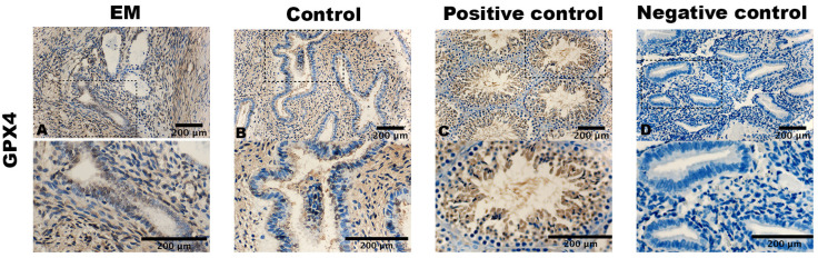
Ferroptosis, an iron-dependent form of non-apoptotic cell death, plays a pivotal role in various diseases and is gaining considerable attention in the realm of endometriosis. Considering the classical pathomechanism theories, we hypothesized that ferroptosis, potentially driven by increased iron content at ectopic sites, may contribute to the progression of endometriosis. This retrospective case-control study provides a comprehensive immunohistochemical assessment of the expression and tissue distribution of established ferroptosis markers: GPX4, ACSL4, and TfR1 in endometriosis patients. The case group consisted of 38 women with laparoscopically and histologically confirmed endometriosis and the control group consisted of 18 women with other gynecological conditions. Our study revealed a significant downregulation of GPX4 in stromal cells of endometriosis patients (M = 59.7% ± 42.4 versus 90.0% ± 17.5 in the control group, t (54) = -2.90, p = 0.005). This finding aligned with slightly, but not significantly, higher iron levels detected in the blood of endometriosis patients, using hemoglobin as an indirect predictor (Hb 12.8 (12.2-13.5) g/dL versus 12.5 (12.2-13.4) g/dL in the control group; t (54) = -0.897, p = 0.374). Interestingly, there was no concurrent upregulation of TfR1 (M = 0.7 ± 1.2 versus 0.2 ± 0.4 for EM, t (54) = 2.552, p = 0.014), responsible for iron uptake into cells. Our empirical findings provide support for the involvement of ferroptosis in the context of endometriosis. However, variances in expression patterns within stromal and epithelial cellular subsets call for further in-depth investigations.
Keywords: ACSL4; GPX4; TfR1; biomarkers; endometriosis; ferroptosis; iron.
Conflict of interest statement
The authors declare no conflicts of interest. The funders had no role in the design of the study; in the collection, analyses, or interpretation of the data; in the writing of the manuscript; or in the decision to publish the results.
Figures


References
-
- Alborzi S., Askary E., Khorami F., Poordast T., Abdulwahid Hashim Alkhalidi B., Hamedi M., Alborzi S., Shahraki H.R. A Detailed Study in Adenomyosis and Endometriosis: Evaluation of the Rate of Coexistence Between Uterine Adenomyosis and DIE According to Imaging and Histopathology Findings. Reprod. Sci. 2021;28:2387–2397. doi: 10.1007/s43032-021-00527-0. - DOI - PubMed
MeSH terms
Substances
Grants and funding
LinkOut - more resources
Full Text Sources
Medical
Miscellaneous
