. 2024 Jul 22;14(7):879.
doi: 10.3390/biom14070879.

Radiation-Induced Endothelial Ferroptosis Accelerates Atherosclerosis via the DDHD2-Mediated Nrf2/GPX4 Pathway

Affiliations

Radiation-Induced Endothelial Ferroptosis Accelerates Atherosclerosis via the DDHD2-Mediated Nrf2/GPX4 Pathway

Xi Su et al. Biomolecules. .

Abstract

This study sought to explore potential roles of endothelial ferroptosis in radiation-associated atherosclerosis (RAA) and molecular mechanisms behind this phenomenon. Here, an in vivo RAA mouse model was used and treated with ferroptosis inhibitors. We found that the RAA group had a higher plaque burden and a reduction in endothelial cells with increased lipid peroxidation compared to the control group, while ameliorated by liproxstatin-1. In vitro experiments further confirmed that radiation induced the occurrence of ferroptosis in human artery endothelial cells (HAECs). Then, proteomics analysis of HAECs identified domain-containing protein 2 (DDHD2) as a co-differentially expressed protein, which was enriched in the lipid metabolism pathway. In addition, the level of lipid peroxidation was elevated in DDHD2-knockdown HAECs. Mechanistically, a significant decrease in the protein and mRNA expression of glutathione peroxidase 4 (GPX4) was observed in HAECs following DDHD2 knockdown. Co-immunoprecipitation assays indicated a potential interaction between DDHD2 and nuclear factor erythroid 2-related factor 2 (Nrf2). The downregulation of Nrf2 protein was also detected in DDHD2-knockdown HAECs. In conclusion, our findings suggest that radiation-induced endothelial ferroptosis accelerates atherosclerosis, and DDHD2 is a potential regulatory protein in radiation-induced endothelial ferroptosis through the Nrf2/GPX4 pathway.

Keywords: DDHD2; Nrf2/GPX4; endothelial injury; ferroptosis; radiation-associated atherosclerosis.


Conflict of interest statement

The authors declare no conflicts of interest.

Figures

Figure 1
Figure 1
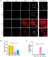
Ferroptosis inhibitor alleviates radiation-induced atherosclerosis lesions. (A) The study design scheme illustrates groups with treatment. (B) The irradiation field in terms of anatomic overview (2 × 1.5 cm2). (C) Representative images of microscopy and sections stained with hematoxylin and eosin, Masson, and Oil Red of the left carotid artery. (D) Quantification of the length of carotid atherosclerotic plaque from ApoE−/− mice in each group (n = 8). (E) Quantification of the oil red positive area of carotid atherosclerotic plaque from ApoE−/− mice in each group (n = 8). **** p < 0.0001.
Figure 2
Figure 2
Ferroptosis inhibitor ameliorates lipid peroxidation and endothelial injury of RAA in ApoE−/− mice. (A) Representative immunofluorescence staining of CD31 (green), 4-HNE (red), and their colocalization (yellow) in carotid atherosclerotic plaques of ApoE−/− mice from each group; the arrows depict the yellow cells/double positive cells. (B) Quantitative analysis of the counts of CD31+ cells in each group (n = 7). (C) Quantitative analysis of the co-expression of CD31+/4-HNE+ cells ratio in each group (n = 7). * p < 0.05; **** p < 0.0001.
Figure 3
Figure 3
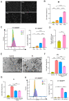
Irradiation promotes HAECs’ death with increased lipid peroxidation. (A) Cell morphology of HAECs 24 h after ionizing radiation (IR). Images were taken at an objective magnification of ×20. (B) Quantity of cell death fraction of HAECs 24 h after irradiation (n = 3) by PI staining. (C) Lipid peroxidation assessment in HAECs irradiated with 0, 4, 8, and 16 Gy after 24 h by C11-BODIPY staining (n = 3). (D) Expression of mRNA PTGS2 in HAECs irradiated with 0 Gy and 16 Gy after 24 h (n = 3). (E) Transmission electron microscopy images showed that HAECs irradiated with 16 Gy (IR) had shrunken mitochondria with enhanced membrane density at 24 h. Black arrow: mitochondria. (F,G) Quantity of cell death fraction of HAECs treated with 0 Gy+DMSO (Control), 16 Gy+DMSO (IR), 16 Gy+2–5 μM Fer-1 (IR+Fer-1), and 16 Gy+10–20 μM DFO (IR+DFO) by PI staining (n = 3). (H) Lipid peroxidation assessment in HAECs treated with 0 Gy+DMSO (control), 0 Gy+Fer-1 (Fer-1), 16 Gy+DMSO (IR), 16 Gy+Fer-1 (IR+Fer-1) after 24 h by C11-BODIPY staining (n = 3). * p < 0.05; ** p < 0.01; *** p < 0.001; **** p < 0.0001.
Figure 4
Figure 4
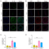
Ferroptosis inhibitor downregulated IL-1β and TNF-α in vivo and in vitro. (A) Representative images of immunofluorescence staining of IL-β (green) in carotid atherosclerotic plaques of ApoE−/− mice. (B) Representative images of immunofluorescence staining of TNF-α (red) in carotid atherosclerotic plaques of ApoE−/− mice. (C,D) Expressions of mRNA IL-1β and TNF-α in HAECs with or without IR and 5 μM Fer-1 treatment at 24 h (n = 4). * p < 0.05; **** p < 0.0001.
Figure 5
Figure 5
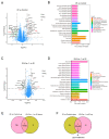
DEPs and KEGG pathway of 4D Label-Free quantitative proteomics analysis. (A) Volcano plot showing the DEPs of IR vs. control group (n = 3/per group). (B) KEGG pathway analysis of DEPs between IR and control group (n = 3/per group). (C) Volcano plot showing the DEPs of IR+Fer-1 vs. IR group (n = 3/per group). (D) KEGG pathway analysis of DEPs between IR+Fer-1 and IR group (n = 3/per group). (E) Venn diagram showing overlap of upregulated DEPs in IR vs. control group and downregulated DEPs in IR+Fer-1 vs. IR group. (F) Venn diagram showing overlap of downregulated DEPs in IR vs. control group and upregulated DEPs in IR+Fer-1 vs. IR group and the involved Reactome pathway of DDHD2 (https://reactome.org/ accessed on 29 May 2023). DEPs: Proteins whose adjusted p-value < 0.05 and |log2Fc|>1.
Figure 6
Figure 6
DDHD2 may be the target of irradiation-induced vascular endothelial ferroptosis. (A) DDHD2 and GAPDH protein levels in HAECs of each group were evaluated by Western blots (n = 3). (B) Western blotting analysis of DDHD2 and GAPDH in HAECs, infected with Ad vector-carrying negative control (shNC) and three different DDHD2-knockdown short hairpin RNA expression plasmids (sh1–3) (n = 4). (C) Expressions of PTGS2 mRNA in shNC; sh1–3 groups were assessed by qRT-PCR. (D) Lipid peroxidation assessment in HAECs infected with sh1–3 and shNC by C11-BODIPY staining (n = 3). (E) GPX4 protein levels in HAECs with shNC, sh1–3 groups, were evaluated by Western blots. (F) Expressions of GPX4 mRNA in shNC; sh1–3 groups were assessed by qRT-PCR (n = 4). (G) Western Blot after co-immunoprecipitation was used to verify the binding of DDHD2 with NRF2. (H) Nrf2 protein levels in HAECs with shNC, sh1–3 groups, evaluated by Western blots. (I) An illustration depicting the role and underlying mechanism of DDHD2 in radiation-induced endothelial ferroptosis: Downregulation of DDHD2 after radiation causes the release of NRF2 from it and promotes NRF2 degradation, inhibiting the NRF2/GPX4 axis and promoting ferroptosis. * p < 0.05; ** p < 0.01; **** p < 0.0001. Original Western blot images are available in Supplementary Materials.

Similar articles

Cited by

References

    1. Stone H.B., Coleman C.N., Anscher M.S., McBride W.H. Effects of radiation on normal tissue: Consequences and mechanisms. Lancet Oncol. 2003;4:529–536. doi: 10.1016/S1470-2045(03)01191-4. - DOI - PubMed
    1. Tocchetti C.G., Ameri P., de Boer R.A., D’Alessandra Y., Russo M., Sorriento D., Ciccarelli M., Kiss B., Bertrand L., Dawson D., et al. Cardiac dysfunction in cancer patients: Beyond direct cardiomyocyte damage of anticancer drugs: Novel cardio-oncology insights from the joint 2019 meeting of the ESC Working Groups of Myocardial Function and Cellular Biology of the Heart. Cardiovasc. Res. 2020;116:1820–1834. doi: 10.1093/cvr/cvaa222. - DOI - PubMed
    1. Shah B.N., Gujral D.M., Chahal N.S., Harrington K.J., Nutting C.M., Senior R. Plaque Neovascularization Is Increased in Human Carotid Atherosclerosis Related to Prior Neck Radiotherapy: A Contrast-Enhanced Ultrasound Study. JACC Cardiovasc. Imaging. 2016;9:668–675. doi: 10.1016/j.jcmg.2015.07.026. - DOI - PubMed
    1. Yamamoto Y., Okawa M., Suzuki K., Tateya I., Yoshimura M., Fushimi Y., Kato E.T., Yoshida K., Miyamoto S. Continuous and Early Progression of Carotid Intima-Media Thickness after Radiotherapy for Head and Neck Cancer: 5-Year Prospective Observational Study. Cerebrovasc. Dis. 2023;52:543–551. doi: 10.1159/000528622. - DOI - PMC - PubMed
    1. Stefan M.F., Herghelegiu C.G., Magda S.L. Accelerated Atherosclerosis and Cardiovascular Toxicity Induced by Radiotherapy in Breast Cancer. Life. 2023;13:1631. doi: 10.3390/life13081631. - DOI - PMC - PubMed

MeSH terms

LinkOut - more resources