Review
. 2024 Jun 24;15(7):832.
doi: 10.3390/genes15070832.

Protein Assemblies in Translesion Synthesis

Affiliations
Review

Protein Assemblies in Translesion Synthesis

Gianluca A Arianna et al. Genes (Basel). .

Abstract

Translesion synthesis (TLS) is a mechanism of DNA damage tolerance utilized by eukaryotic cells to replicate DNA across lesions that impede the high-fidelity replication machinery. In TLS, a series of specialized DNA polymerases are employed, which recognize specific DNA lesions, insert nucleotides across the damage, and extend the distorted primer-template. This allows cells to preserve genetic integrity at the cost of mutations. In humans, TLS enzymes include the Y-family, inserter polymerases, Polη, Polι, Polκ, Rev1, and the B-family extender polymerase Polζ, while in S. cerevisiae only Polη, Rev1, and Polζ are present. To bypass DNA lesions, TLS polymerases cooperate, assembling into a complex on the eukaryotic sliding clamp, PCNA, termed the TLS mutasome. The mutasome assembly is contingent on protein-protein interactions (PPIs) between the modular domains and subunits of TLS enzymes, and their interactions with PCNA and DNA. While the structural mechanisms of DNA lesion bypass by the TLS polymerases and PPIs of their individual modules are well understood, the mechanisms by which they cooperate in the context of TLS complexes have remained elusive. This review focuses on structural studies of TLS polymerases and describes the case of TLS holoenzyme assemblies in action emerging from recent high-resolution Cryo-EM studies.

Keywords: DNA damage tolerance; DNA repair; protein assemblies; protein structure; protein–protein interactions; translesion synthesis.


Conflict of interest statement

The authors declare no conflicts of interest.

Figures

Figure 4
Figure 4
Protein-protein interactions in TLS. (A) Hs PCNA with a Polη PIP-box peptide bound near the interdomain connector loop (IDCL) [130]. Ubiquitin (gray) is ligated at K164 of PCNA by the Rad6/Rad18 E2/E3 pair. A close-up view of the PCNA monomer showing a “three-pronged fork” of Polη PIP (L704, F707, F708) inserting into a hydrophobic pocket in the IDCL region of PCNA, and M701 binding to the Q-pocket. (B) PIP-box and RIR sequences in the human TLS polymerases aligned to their minimal motifs. (C) Hs Rev1 UBM2 bound to a conserved site on ubiquitin centered at L8, I44, and V70 [135]. (D) A model of Hs Polη UBZ bound to ubiquitin (based on the Rad18-UBZ/ubiquitin complex) [132,137]. (E) Hs Polκ RIR motif bound to Rev1-CT with F567 and F568 inserted in a hydrophobic pocket in the N-terminal part of the domain [85]. (F) Ternary complex of Hs Rev7, Rev3 RBM1, and Rev1-CT [83]. Rev7 uses a C-terminal ‘safety-belt’ loop (blue) to latch the Rev3 RBM1 peptide to the core. The C-terminus of the ‘safety-belt’ region forms a β8′-β8′’ hairpin, which extends the core β-sheet (β4-6) and provides the binding interface for Rev1-CT.
Figure 6
Figure 6
Cryo-EM structures of the Sc Polζ and Sc Rev1/Polζ complex. (A) “Daisy-chain” or ring-like architecture of the five-subunit Sc Polζ holoenzyme on p/t DNA. Rev3 accommodates p/t DNA (red) and makes contacts to Pol31, Rev7A, and Rev7B but not Pol32 [97]. Rotation of the structure shows an unstructured C-terminus of Pol32, which contains a PIP-box motif, reaching a tentative PCNA location. (B) An unusual head-to-tail Sc Rev7 dimer within Polζ (top) that does not utilize the canonical HORMA dimerization interface [97]. For comparison, a head-to-head dimer is shown (bottom) formed by Hs Rev7 within the shielding complex [149]. Here the heterodimer of closed (C-Rev7) and open (O-Rev7) protomers is formed via the interface centered at helix αC. (C) Partial structure of the Sc Rev1/Polζ complex in which the Sc Polζ holoenzyme is engaged with p/t DNA [100]. Only the Rev1 CT, BRCT, and M1 regions are visible. The PCNA ring is modeled based on the structure of Polδ/PCNA complex [87]. A close-up view of the Rev1-M1 and Rev1-BRCT interactions with p/t DNA is shown on the right [100].
Figure 1
Figure 1
TLS DNA polymerases in humans and yeast. (A,B) Domains and subunits of (A) human and (B) yeast Y-family polymerases Polη, Polι, Polκ, Rev1, and B-family polymerase Pol ζ. The core catalytic domain of each Y-family polymerase (including fingers, thumb, and palm subdomains) is followed by a polymerase associated domain (PAD). B-family Polζ lacks the PAD domain but includes an inactive endonuclease domain within the catalytic core. The binding modules of Y-family TLS polymerases include PCNA-interacting PIP and Rev1-interacting RIR motifs, ubiquitin-binding UBM (Rev1, Polι) and UBZ (Polη, Polκ) domains, Rev1 N-terminal BRCT and the C-terminal CT domains, and the N-terminal α-helical region of Rev1 termed the “M1” motif. Polη includes a nuclear localization signal (NLS). Polζ comprises four subunits: Rev3, Rev7, PolD2/PolD3 (humans), or Pol31/Pol32 (yeast). The Rev3 subunit of Polζ harbors a catalytic core domain, an N-terminal domain (NTD), a positively charged domain (PCD, humans), two Rev7-binding motifs (RBMs), and a C-terminal domain (CTD) interacting with the PolD2/PolD3 or Pol31/Pol32 module. (C) Schematic of Rev1/Polζ-dependent TLS mutasome assembled on ubiquitinated PCNA, including an ‘inserter’ TLS polymerase Polη, and ‘extender’ TLS polymerase Polζ and a scaffold protein Rev1. Possible interactions between domains and subunits of the TLS proteins are indicated. Adapted with permission from [32].
Figure 2
Figure 2
Catalytic domains of human Y-family TLS DNA polymerases. Each Y-family polymerase features fingers (blue), palm (red), thumb (green), and PAD (yellow) domains. Insets for A–D show close-ups of polymerase active sites in the process of nucleotide insertion. (A) Hs Polη bypassing a cisplatin DNA adduct. (B) Polκ bypassing benzo[a]pyrene adduct. (C) Hs Polι bypassing 8-oxoG lesion. (D) Hs Rev1 inserting cytosine on a normal p/t DNA.
Figure 3
Figure 3
Cryo-EM structure of the Sc Polζ catalytic domain. (A) The Sc Rev3 catalytic domain on p/t DNA, including the fingers (yellow), thumb (orange), palm (cyan), NTD (blue), and inactive exonuclease (purple) domains [97]. The CysB domain within Rev3 CTD that binds Pol31 is also shown. The inset shows a close-up of the Rev3 active site, revealing the basis for Polζ intolerance to mismatches during nucleotide insertion. Residues from the fingers (yellow) and palm (blue) domains make extensive contacts with the nascent base pair (red). (B) Structural basis for Polζ extension past DNA mismatches [97]. Due to contacts with αxC and palm-loop, the NTD-palm linker in the Rev3 active site is held further away from DNA than in Pol3. (C) Structural basis of the impaired exonuclease activity of Polζ [97]. Unlike Pol3, Rev3 lacks the β-hairpin used to pass mismatched DNA from the active site to the exonuclease domain.
Figure 5
Figure 5
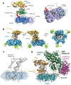
Cryo-EM structure of the human Polκ holoenzyme. (A) Polκ bound to p/t DNA (gray) and the front face of unmodified PCNA (blue). The catalytic core of Polκ comprises the palm (tan), fingers (yellow), thumb (orange), N-clasp (pink), and PAD domains (green). An ‘inverting’ helix C-terminal to PAD orients the internal Polκ PIP-box motif (red) towards the binding site on the PCNA1 protomer. The right panel shows a structural comparison of the Hs Polκ PIP1 and Hs Polδ PIP motifs bound to a PCNA protomer. (B) Polκ in complex with p/t DNA and mono-ubiquitinated PCNA with ubiquitin moieties visible on the PCNA back face. (C) DNA that exits the Polκ holoenzyme tilts 47° relative to the PCNA axis, resulting in DNA bending when passing through the PCNA ring. (D) A model of a PCNA ‘tool belt’ that simultaneously binds to the Polκ/DNA (yellow/gray) complex and apo Polδ (green/magenta). Adapted with permission from [96].

Similar articles

Cited by

  • Targeting the DNA damage response in cancer.
    Federica G, Michela C, Giovanna D. Federica G, et al. MedComm (2020). 2024 Oct 31;5(11):e788. doi: 10.1002/mco2.788. eCollection 2024 Nov. MedComm (2020). 2024. PMID: 39492835 Free PMC article. Review.

References

    1. Friedberg E.C., Wood R.D., Walker G.C., Schultz R.A., Slede W., Ellenberger T. DNA Repair and Mutagenesis. 2nd ed. ASM Press; Washington, DC, USA: 2006.
    1. De Bont R., van Larebeke N. Endogenous DNA damage in humans: A review of quantitative data. Mutagenesis. 2004;19:169–185. doi: 10.1093/mutage/geh025. - DOI - PubMed
    1. Bjelland S., Seeberg E. Mutagenicity, toxicity and repair of DNA base damage induced by oxidation. Mutat. Res. 2003;531:37–80. doi: 10.1016/j.mrfmmm.2003.07.002. - DOI - PubMed
    1. Davies R.J.H. Ultraviolet radiation damage in DNA. Biochem. Soc. Trans. 1995;23:407–418. doi: 10.1042/bst0230407. - DOI - PubMed
    1. Chatterjee N., Walker G.C. Mechanisms of DNA damage, repair, and mutagenesis. Env. Mol. Mutagen. 2017;58:235–263. doi: 10.1002/em.22087. - DOI - PMC - PubMed

MeSH terms

LinkOut - more resources